Your shopping cart is empty!
")
| Reactivity: | Human, Mouse, Rat |
| Applications: | WB, IHC, IF/IC, ELISA |
| Host Species: | Rabbit |
| Isotype: | IgG |
| Clonality: | Monoclonal antibody |
| Gene Name: | superoxide dismutase 2 |
| Gene Symbol: | SOD2 |
| Synonyms: | GC1; IPOB; IPO-B; MNSOD; MVCD6; GClnc1; Mn-SOD; D2 |
| Gene ID: | 6648 |
| UniProt ID: | P04179 |
| Clone ID: | 9T2Z1 |
| Immunogen: | A synthetic peptide corresponding to a sequence within amino acids 1-100 of human SOD2 (P04179). |
| Dilution: | WB 1:2000-1:8000; IHC 1:200-1:2000; IF/IC 1:200-1:800 |
| Purification Method: | Affinity purification |
| Concentration: | 0.25 mg/mL |
| Buffer: | PBS with 0.02% sodium azide, 0.05% BSA, 50% glycerol, pH7.3. |
| Storage: | Store at -20°C. Avoid freeze / thaw cycles. |
| Documents: | Manual-SOD2 monoclonal antibody |
Background
This gene is a member of the iron/manganese superoxide dismutase family. It encodes a mitochondrial protein that forms a homotetramer and binds one manganese ion per subunit. This protein binds to the superoxide byproducts of oxidative phosphorylation and converts them to hydrogen peroxide and diatomic oxygen. Mutations in this gene have been associated with idiopathic cardiomyopathy (IDC), premature aging, sporadic motor neuron disease, and cancer. Alternative splicing of this gene results in multiple transcript variants. A related pseudogene has been identified on chromosome 1.
Images
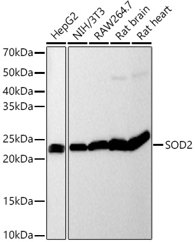 | Western blot analysis of various lysates, using SOD2 Rabbit mAb (A19576) at 1:2000 dilution. Secondary antibody: HRP-conjugated Goat anti-Rabbit IgG (H+L) (AS014) at 1:10000 dilution. Lysates/proteins: 25μg per lane. Blocking buffer: 3% nonfat dry milk in TBST. Detection: ECL Basic Kit (RM00020). Exposure time: 20s. |
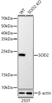 | Western blot analysis of lysates from wild type(WT) and SOD2 knockout (KO) 293T(KO) cells, using SOD2 Rabbit mAb (A19576) at 1:2000 dilution. Secondary antibody: HRP-conjugated Goat anti-Rabbit IgG (H+L) (AS014) at 1:10000 dilution. Lysates/proteins: 25μg per lane. Blocking buffer: 3% nonfat dry milk in TBST. Detection: ECL Basic Kit (RM00020). Exposure time: 20s. |
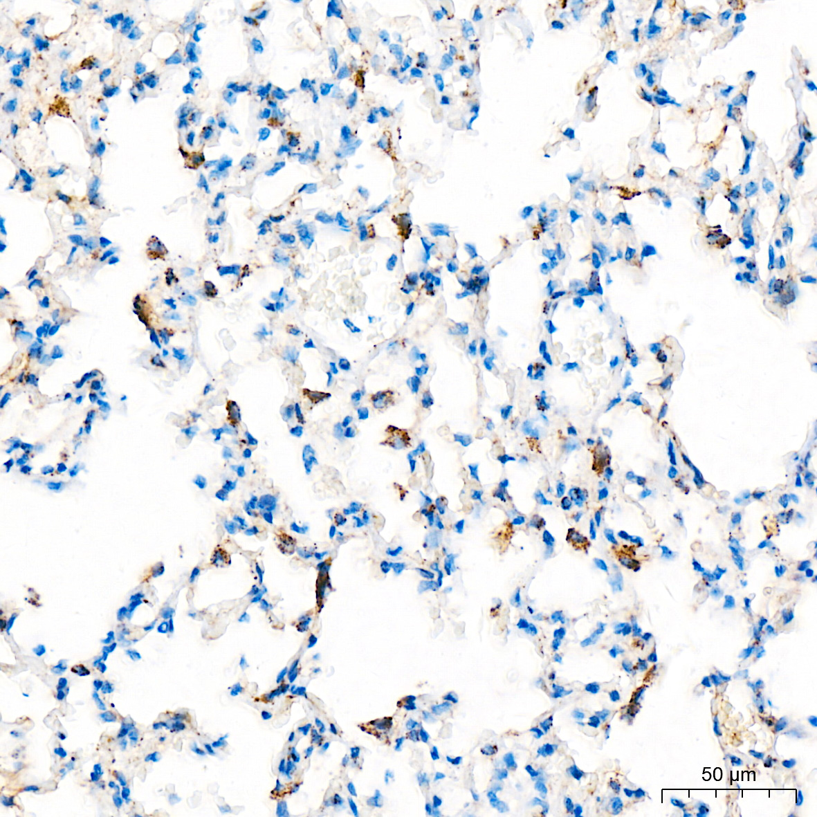 | Immunohistochemistry analysis of paraffin-embedded Rat lung tissue using [KO Validated] SOD2 Rabbit mAb (A19576) at a dilution of 1:200 (40x lens). High pressure antigen retrieval performed with 0.01M Citrate Buffer (pH 6.0) prior to IHC staining. |
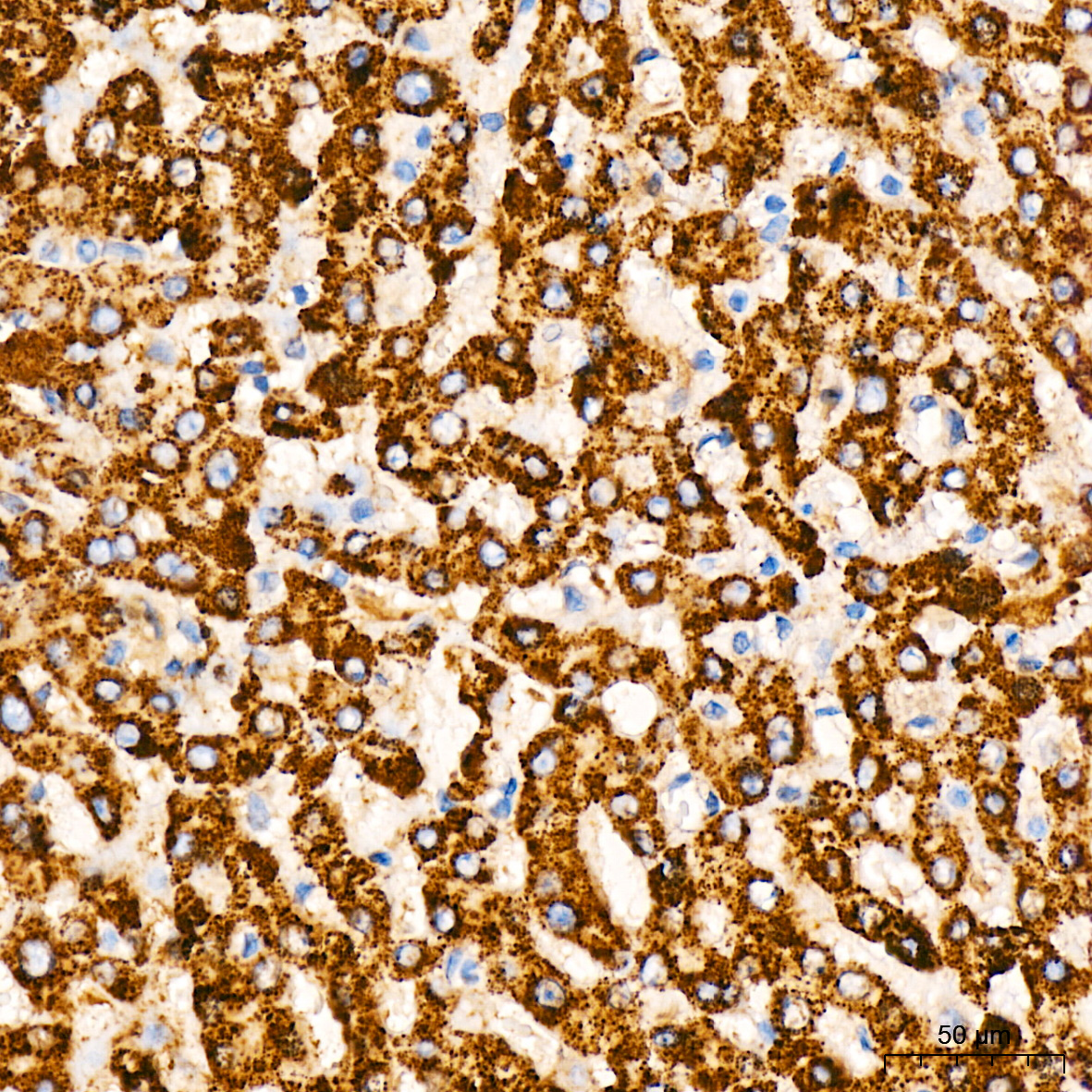 | Immunohistochemistry analysis of paraffin-embedded Human liver tissue using [KO Validated] SOD2 Rabbit mAb (A19576) at a dilution of 1:200 (40x lens). High pressure antigen retrieval performed with 0.01M Citrate Buffer (pH 6.0) prior to IHC staining. |
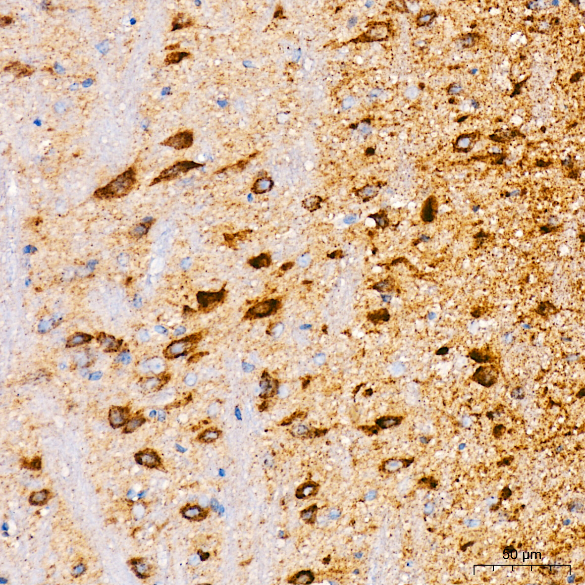 | Immunohistochemistry analysis of paraffin-embedded Mouse brain tissue using [KO Validated] SOD2 Rabbit mAb (A19576) at a dilution of 1:200 (40x lens). High pressure antigen retrieval performed with 0.01M Citrate Buffer (pH 6.0) prior to IHC staining. |
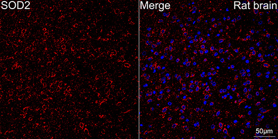 | Confocal imaging of paraffin-embedded Rat brain tissue using [KO Validated] SOD2 Rabbit mAb (A19576, dilution 1:200) followed by a further incubation with Cy3 Goat Anti-Rabbit IgG (H+L) (AS007, dilution 1:500) (Red). DAPI was used for nuclear staining (Blue). High pressure antigen retrieval performed with 0.01M Citrate Buffer (pH 6.0) prior to IF staining. Objective: 40x. |
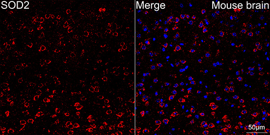 | Confocal imaging of paraffin-embedded Mouse brain tissue using [KO Validated] SOD2 Rabbit mAb (A19576, dilution 1:200) followed by a further incubation with Cy3 Goat Anti-Rabbit IgG (H+L) (AS007, dilution 1:500)(Red). DAPI was used for nuclear staining (Blue). High pressure antigen retrieval performed with 0.01M Citrate Buffer (pH 6.0) prior to IF staining. Objective: 40x. |
You may also be interested in:

