Your shopping cart is empty!
")
| Reactivity: | Human, Mouse, Rat |
| Applications: | WB, IF/IC, IP, ELISA |
| Host Species: | Rabbit |
| Isotype: | IgG |
| Clonality: | Monoclonal antibody |
| Gene Name: | SRC proto-oncogene, non-receptor tyrosine kinase |
| Gene Symbol: | SRC |
| Synonyms: | ASV; SRC1; THC6; c-SRC; p60-Src; rc |
| Gene ID: | 6714 |
| UniProt ID: | P12931 |
| Clone ID: | 7G6M9 |
| Immunogen: | A synthetic peptide corresponding to a sequence within amino acids 1-100 of human Src (P12931). |
| Dilution: | WB 1:1000-1:2000; IF/IC 1:100-1:400 |
| Purification Method: | Affinity purification |
| Concentration: | 0.5 mg/mL |
| Buffer: | PBS with 0.02% sodium azide, 0.05% BSA, 50% glycerol, pH7.3. |
| Storage: | Store at -20°C. Avoid freeze / thaw cycles. |
| Documents: | Manual-SRC monoclonal antibody |
Background
This gene is highly similar to the v-src gene of Rous sarcoma virus. This proto-oncogene may play a role in the regulation of embryonic development and cell growth. The protein encoded by this gene is a tyrosine-protein kinase whose activity can be inhibited by phosphorylation by c-SRC kinase. Mutations in this gene could be involved in the malignant progression of colon cancer. Two transcript variants encoding the same protein have been found for this gene.
Images
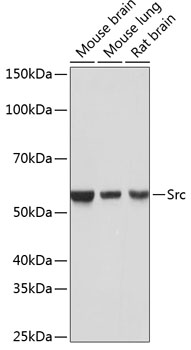 | Western blot analysis of various lysates using [KO Validated] Src Rabbit mAb (A19119) at 1:1000 dilution. Secondary antibody: HRP-conjugated Goat anti-Rabbit IgG (H+L) (AS014) at 1:10000 dilution. Lysates/proteins: 25μg per lane. Blocking buffer: 3% nonfat dry milk in TBST. Detection: ECL Basic Kit (RM00020). Exposure time: 1s. |
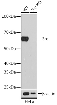 | Western blot analysis of lysates from wild type (WT) and Src knockout (KO) HeLa cells, using [KO Validated] Src Rabbit mAb (A19119) at 1:1000 dilution. Secondary antibody: HRP-conjugated Goat anti-Rabbit IgG (H+L) (AS014) at 1:10000 dilution. Lysates/proteins: 25μg per lane. Blocking buffer: 3% nonfat dry milk in TBST. Detection: ECL Basic Kit (RM00020). Exposure time: 1min. |
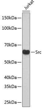 | Western blot analysis of lysates from Jurkat cells, using [KO Validated] Src Rabbit mAb (A19119) at 1:1000 dilution. Secondary antibody: HRP-conjugated Goat anti-Rabbit IgG (H+L) (AS014) at 1:10000 dilution. Lysates/proteins: 25μg per lane. Blocking buffer: 3% nonfat dry milk in TBST. Detection: ECL Basic Kit (RM00020). Exposure time: 1min. |
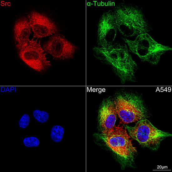 | Confocal imaging of A549 cells using [KO Validated] Src Rabbit mAb (A19119,at dilution of 1:100) (Red). The cells were counterstained with α-Tubulin Mouse mAb (AC012,dilution 1:400) (Green). DAPI was used for nuclear staining (blue). Objective: 100x. |
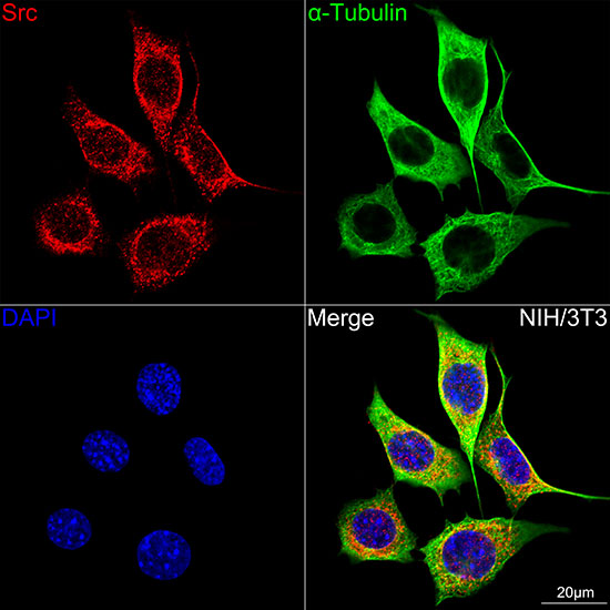 | Confocal imaging of NIH/3T3 cells using [KO Validated] Src Rabbit mAb (A19119,at dilution of 1:100) (Red). The cells were counterstained with α-Tubulin Mouse mAb (AC012,dilution 1:400) (Green). DAPI was used for nuclear staining (blue). Objective: 100x. |
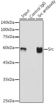 | Immunoprecipitation analysis of 600 μg extracts of Mouse brain using 3 μg Src antibody (A19119). Western blot was performed from the immunoprecipitate using Src antibody (A19119) at a dilution of 1:1000. |
You may also be interested in:

