LiFluor™ 488 EU Imaging Kit provides a simple and robust assay for detection of global RNA transcription temporally and spatially in cells and tissues. The ability to detect newly synthesized RNA or changes in RNA levels resulting from disease, environmental damage or drug treatments is an important aspect of toxicological profiling. In this assay, newly synthesized RNA can be detected in a simple, two-step procedure. In step one, the alkyne-modified nucleoside, 5-ethynyl uridine (EU), is fed to cells or animals and actively incorporated into nascent RNA. The small size of the tag enables efficient incorporation of the modified nucleoside into RNA, but not into DNA. Detection utilizes the “click" reaction between an azide and an alkyne where the modified RNA is detected with a corresponding azide-containing dye. The assay is compatible with antibody-based detection for multiplex imaging.
Key Features
• Simple: Labeling is complete in two steps.
• Fast: Results in as little as 90 minutes.
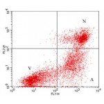
• Multiplex: Compatible with cell cycle dyes and antibody-based stains.
Specifications
1. Platform: Fluorescence Microscope
2. Detection Method: Fluorescent
3. Ex/Em: 495/520 nm
Applications
Cell proliferation analysis in vitro and in vivo.
Components
1. EU: 1 ml
2. LiFluor 488 azide: 100 µl
3. EU reaction buffer: 50 ml
4. CuSO4: 1 ml
5. EdU buffer additive: 200 mg
6. Hoechst 33342: 70 µl
Storage
Store at -20°C and protect from light.
Case Study

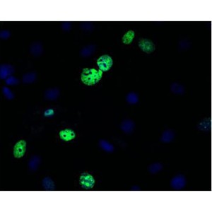
Detection of newly synthesized RNA in cell. A549 cells were treated with 100 µM EU for 2 hr, then detected with LiFluor™ 488 azide (green), cells were counterstained with DAPI (blue).

Detection of newly synthesized RNA in mouse tissue. A mouse was injected intraperitoneally with 50 mg EU per kilogram body weight, then sacrificed at 0, 12, and 24 hr. The mouse intestine tissue was detected with LiFluor™ 488 azide (green), and counterstained with DAPI (blue).
Download


