Internucleosomal cleavage of DNA is a hallmark of apoptosis. DNA cleavage in apoptotic cells can be detected in situ in fixed cells or tissue sections using the terminal deoxynucleotidyl transferase (TdT) mediated dUTP nick-end labeling (TUNEL) method. TUNEL is highly selective for the detection of apoptotic cells but not necrotic cells or cells with DNA strand breaks resulting from irradiation or drug treatment.
In the TUNEL assay, TdT enzyme catalyzes the addition of labeled dUTP to the 3’ ends of cleaved DNA fragments. Hapten-tagged dUTP (e.g. digoxigenin-dUTP or biotin-dUTP) can be detected using secondary reagents (e.g. anti-digoxigenin antibodies or streptavidin) for fluorescence or colorimetric detection. Alternatively, fluorescent dye-conjugated dUTP can be used for direct detection of fragmented DNA by fluorescence microscopy or flow cytometry. The TUNEL LiFluor™ 594 Apoptosis Detection Kit contains dUTP conjugated to biotin and Streptavidin conjugated to bright and photostable LiFluor™ 594 red fluorescent dye, for bright fluorescent TUNEL staining.
Key Features
• Broad application: Suitable for FFPE tissue, frozen tissue and cells.
• High sensitivity: Can detect a low population of apoptotic cells.
• Bright apoptotic signal: Uses LiFluor™ dyes, resulting in a stable, non-photobleaching fluorescent signal.
Specifications
1. Platform: Fluorescence Microscopy
2. Detection Method: Fluorescent
3. Ex/Em: 590/615 nm
Applications
Cell apoptosis analysis
Component
1. TdT reaction buffer: 8 ml
2. TdT enzyme: 100 µl
3. Biotin-11-dUTP: 50 µl
4. LiFluor 594-Streptavidin: 50 µl
5. DNase I: 10 µl
6. DNase I buffer: 1 ml
7. Proteinase K: 50 µl
Storage
Store at -20°C and protect from light.
Case Study

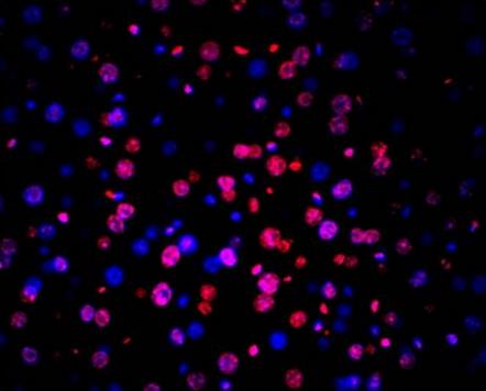
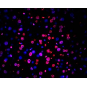
Detection of apoptotic cells in mouse tongue tissue using TUNEL LiFluor™ 594 Apoptosis Detection Kit.


