The LiFluor™ EdU 647 Flow Cytometry Assay Kit presents a more straightforward and robust method for analyzing DNA replication in proliferating cells, offering a significant advancement over the traditional BrdU cell proliferation assay. This assay employs EdU, a modified thymidine analogue, which is effectively integrated into the DNA of newly synthesized cells. It is then labeled with a vivid, photostable LiFluor™ 647 dye through a rapid and highly-specific click reaction. The fluorescent tagging of proliferating cells is both precise and compatible with antibody methods, a result of the mild click protocol employed. The analysis of newly synthesized DNA is performed using the 633 nm laser line of the flow cytometer, ensuring a high level of accuracy and efficiency in detection.
Key Features
• Simple: Labeling is complete in two steps.
• Accurate: Superior results compared to BrdU assay.
• Fast: Results in as little as 90 minutes.
• Multiplex: Compatible with cell cycle dyes and antibody-based stain.
Specifications
1. Platform: Fluorescence Microscope
2. Detection Method: Fluorescent
3. Ex/Em: 650/665 nm
Applications
Cell proliferation analysis by flow cytometer
Components
1. EdU: 2×1 ml
2. LiFluor 647 azide: 150 µl
3. LiFluor fixative: 5 ml
4. Permeabilization and wash reagent: 50 ml
5. CuSO4: 1 ml
6. EdU buffer additive: 200 mg
Storage
Store at -20°C and protect from light.
Case Study

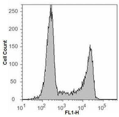
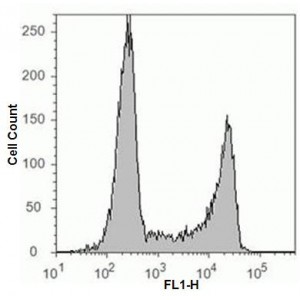
Cell proliferation analysis with LiFluor™ 647 EdU Flow Cytometry Assay Kit. Jurkat cells were treated with 10 µM EdU for 2 hours and detected according to the recommended staining protocol. The figures show a clear separation of proliferating cells which have incorporated EdU and nonproliferating cells which have not.


