Your shopping cart is empty!
")
| Reactivity: | Human, Mouse, Rat |
| Applications: | WB, IHC, ELISA |
| Host Species: | Rabbit |
| Isotype: | IgG |
| Clonality: | Monoclonal antibody |
| Gene Name: | deoxycytidine kinase |
| Gene Symbol: | DCK |
| Synonyms: | dCK |
| Gene ID: | 1633 |
| UniProt ID: | P27707 |
| Clone ID: | 5Q7D0 |
| Immunogen: | Recombinant fusion protein containing a sequence corresponding to amino acids 1-260 of human DCK (NP_000779.1). |
| Dilution: | WB 1:500-1:1000; IHC 1:100-1:500 |
| Purification Method: | Affinity purification |
| Concentration: | 1.88 mg/ml |
| Buffer: | PBS with 0.05% proclin300, 0.05% BSA, 50% glycerol, pH7.3. |
| Storage: | Store at -20°C. Avoid freeze / thaw cycles. |
| Documents: | Manual-DCK monoclonal antibody |
Background
Deoxycytidine kinase (DCK) is required for the phosphorylation of several deoxyribonucleosides and their nucleoside analogs. Deficiency of DCK is associated with resistance to antiviral and anticancer chemotherapeutic agents. Conversely, increased deoxycytidine kinase activity is associated with increased activation of these compounds to cytotoxic nucleoside triphosphate derivatives. DCK is clinically important because of its relationship to drug resistance and sensitivity.
Images
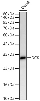 | Western blot analysis of lysates from Daudi cells using [KD Validated] DCK Rabbit mAb (A24071) at 1:1000 dilution. Secondary antibody: HRP-conjugated Goat anti-Rabbit IgG (H+L) (AS014) at 1:10000 dilution. Lysates/proteins: 25 μg per lane. Blocking buffer: 3% nonfat dry milk in TBST. Detection: ECL Basic Kit (RM00020). Exposure time:60s. |
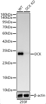 | Western blot analysis of lysates from wild type (WT) and DCK knockdown (KD) 293F cells using [KD Validated] DCK Rabbit mAb (A24071) at 1:1000 dilution. Secondary antibody:HRP Goat Anti-Rabbit IgG (H+L)(AS014) at 1:10000 dilution.Lysates/proteins: 25 μg per lane. Blocking buffer: 3% nonfat dry milk in TBST. Detection:ECL Basic Kit (RM00020). Exposuretime: 60s. |
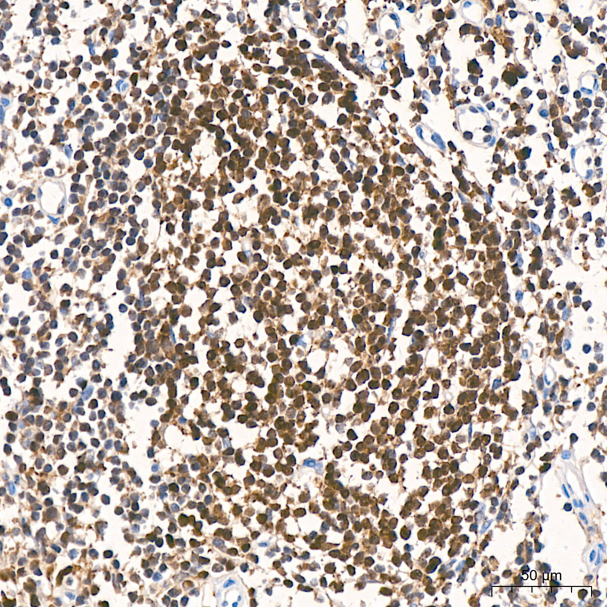 | Immunohistochemistry analysis of paraffin-embedded Human tonsil tissue using [KD Validated] DCK Rabbit mAb (A24071) at a dilution of 1:300 (40x lens). High pressure antigen retrieval performed with 0.01M Citrate Bufferr (pH 6.0) prior to IHC staining. |
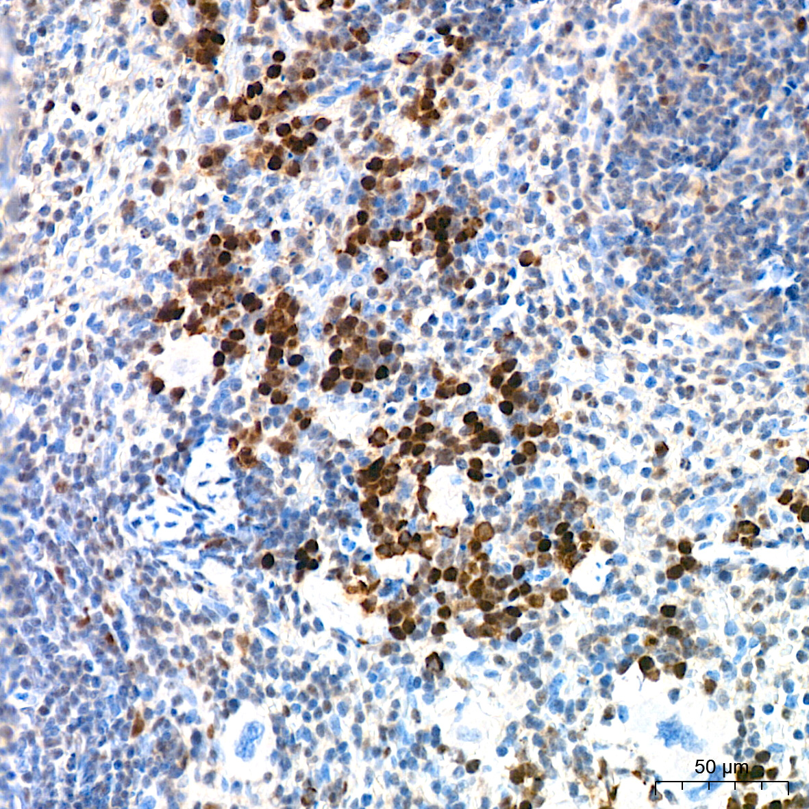 | Immunohistochemistry analysis of paraffin-embedded Mouse spleen tissue using [KD Validated] DCK Rabbit mAb (A24071) at a dilution of 1:300 (40x lens). High pressure antigen retrieval performed with 0.01M Citrate Bufferr (pH 6.0) prior to IHC staining. |
 | Immunohistochemistry analysis of paraffin-embedded Rat spleen tissue using [KD Validated] DCK Rabbit mAb (A24071) at a dilution of 1:300 (40x lens). High pressure antigen retrieval performed with 0.01M Citrate Bufferr (pH 6.0) prior to IHC staining. |
You may also be interested in:

