Your shopping cart is empty!
")
KD-Validated HSP90B1 Rabbit mAb (20 μl)
| Reactivity: | Human, Mouse, Rat |
| Applications: | WB, IHC, ELISA |
| Host Species: | Rabbit |
| Isotype: | IgG |
| Clonality: | Monoclonal antibody |
| Gene Name: | heat shock protein 90 beta family member 1 |
| Gene Symbol: | HSP90B1 |
| Synonyms: | ECGP; GP96; TRA1; GRP94; HEL35; HEL-S-125m |
| Gene ID: | 7184 |
| UniProt ID: | P14625 |
| Clone ID: | 7P4V7 |
| Immunogen: | A synthetic peptide corresponding to a sequence within amino acids 616-715 of human HSP90B1 (NP_003290.1). |
| Dilution: | WB 1:1000-1:4000; IHC 1:200-1:800 |
| Purification Method: | Affinity purification |
| Concentration: | 1 mg/mL |
| Buffer: | PBS with 0.05% proclin300, 0.05% BSA, 50% glycerol, pH7.3. |
| Storage: | Store at -20°C. Avoid freeze / thaw cycles. |
| Documents: | Manual-HSP90B1 monoclonal antibody |
Background
This gene encodes a member of a family of adenosine triphosphate(ATP)-metabolizing molecular chaperones with roles in stabilizing and folding other proteins. The encoded protein is localized to melanosomes and the endoplasmic reticulum. Expression of this protein is associated with a variety of pathogenic states, including tumor formation. There is a microRNA gene located within the 5' exon of this gene. There are pseudogenes for this gene on chromosomes 1 and 15.
Images
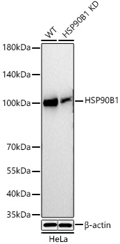
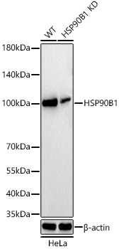 | Western blot analysis of lysates from wild type (WT) and HSP90B1 knockdown (KD) HeLa cells using [KD Validated] HSP90B1 Rabbit mAb (A26311) at 1:2000 dilution incubated overnight at 4℃. Secondary antibody: HRP-conjugated Goat anti-Rabbit IgG (H+L) (AS014) at 1:10000 dilution. Lysates/proteins: 25 μg per lane. Blocking buffer: 3% nonfat dry milk in TBST. Detection: ECL Basic Kit (RM00020). Exposure time: 20s. |
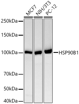 | Western blot analysis of various lysates using [KD Validated] HSP90B1 Rabbit mAb (A26311) at 1:2000 dilution incubated overnight at 4℃. Secondary antibody: HRP-conjugated Goat anti-Rabbit IgG (H+L) (AS014) at 1:10000 dilution. Lysates/proteins: 25 μg per lane. Blocking buffer: 3% nonfat dry milk in TBST. Detection: ECL Basic Kit (RM00020). Exposure time: 60s. |
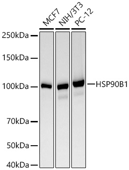
 | Western blot analysis of various lysates using [KD Validated] HSP90B1 Rabbit mAb (A26311) at 1:2000 dilution incubated overnight at 4℃. Secondary antibody: HRP-conjugated Goat anti-Rabbit IgG (H+L) (AS014) at 1:10000 dilution. Lysates/proteins: 25 μg per lane. Blocking buffer: 3% nonfat dry milk in TBST. Detection: ECL Basic Kit (RM00020). Exposure time: 60s. |
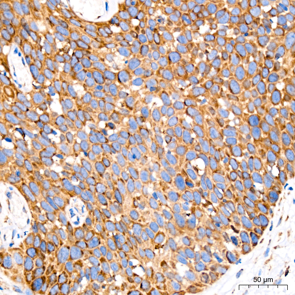
 | Immunohistochemistry analysis of paraffin-embedded Human cervix cancer tissue using [KD Validated] HSP90B1 Rabbit mAb (A26311) at a dilution of 1:200 (40x lens). High pressure antigen retrieval performed with 0.01M Citrate Buffer (pH 6.0) prior to IHC staining. |
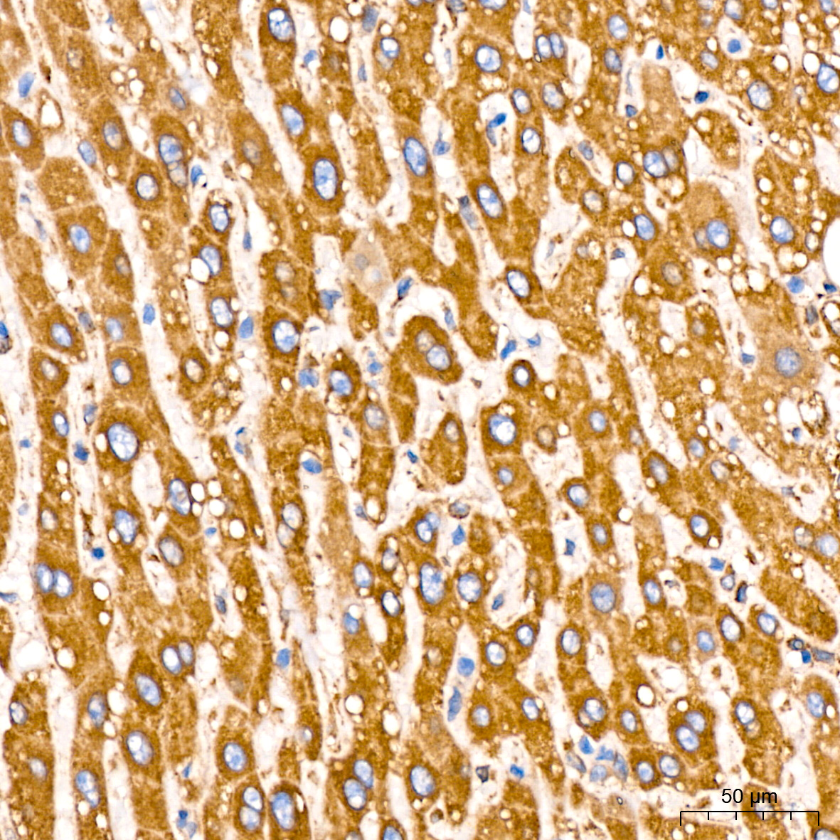
 | Immunohistochemistry analysis of paraffin-embedded Human liver tissue using [KD Validated] HSP90B1 Rabbit mAb (A26311) at a dilution of 1:200 (40x lens). High pressure antigen retrieval performed with 0.01M Citrate Buffer (pH 6.0) prior to IHC staining. |
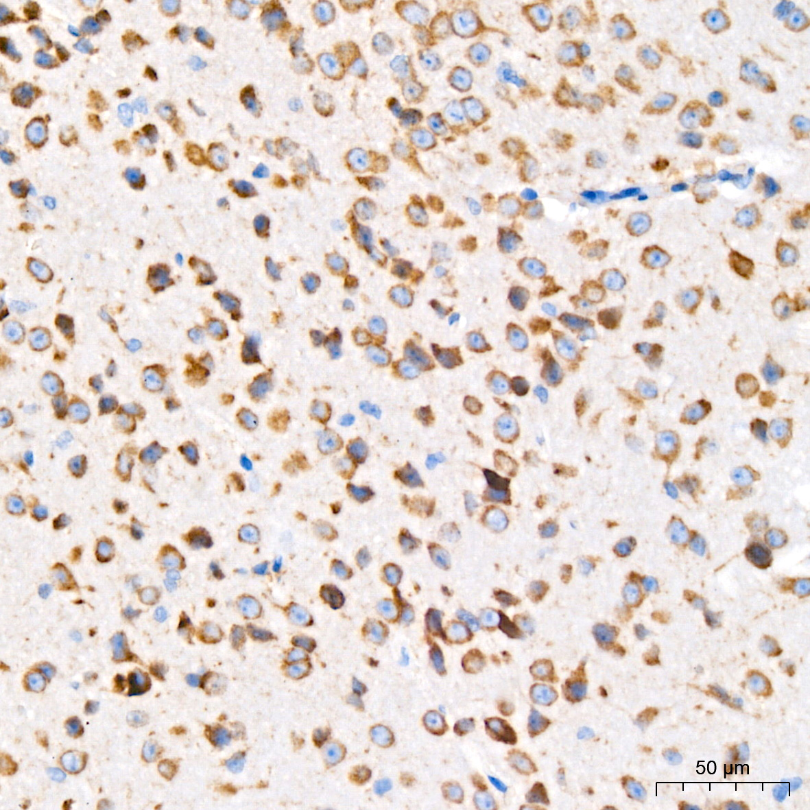
 | Immunohistochemistry analysis of paraffin-embedded Mouse brain tissue using [KD Validated] HSP90B1 Rabbit mAb (A26311) at a dilution of 1:200 (40x lens). High pressure antigen retrieval performed with 0.01M Citrate Buffer (pH 6.0) prior to IHC staining. |
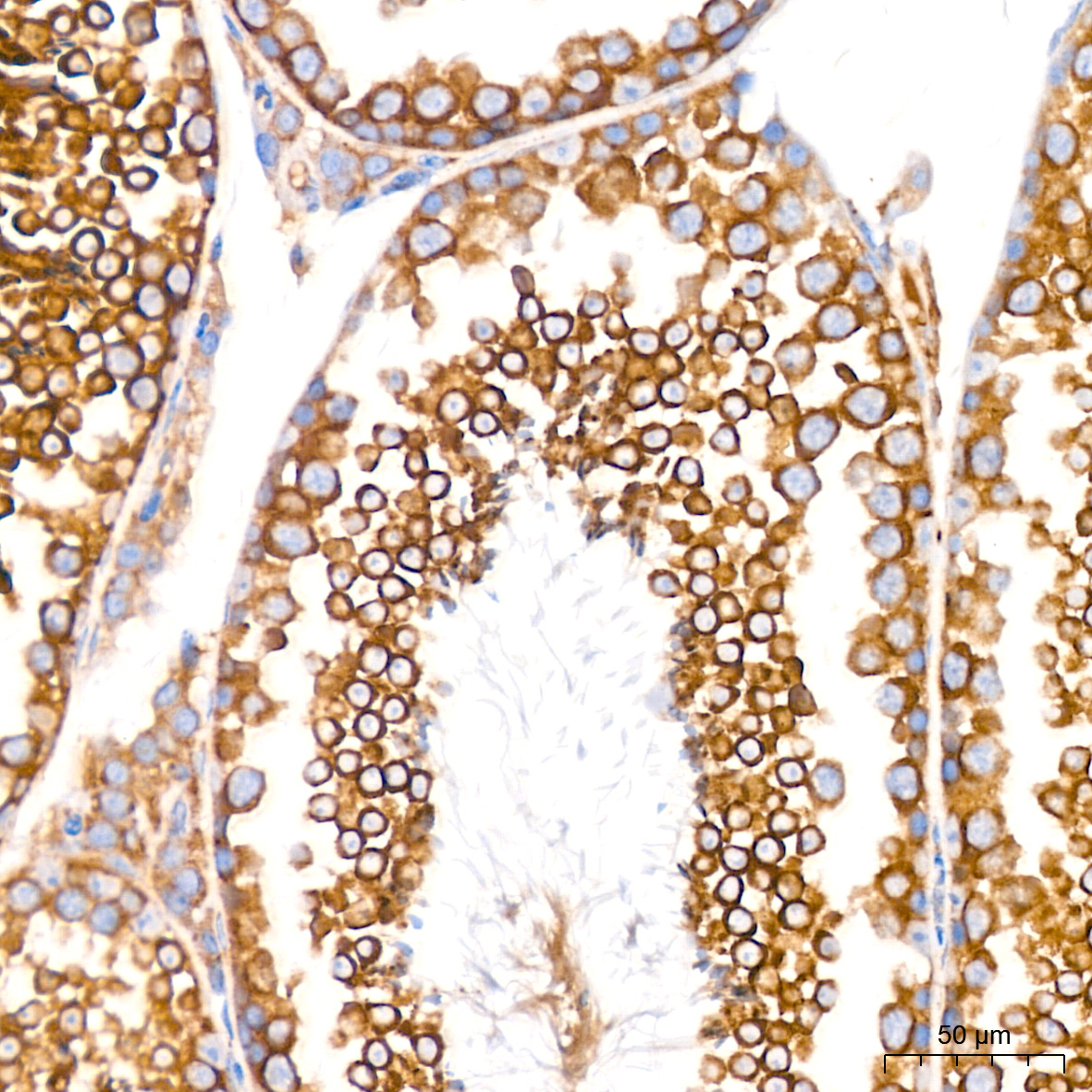
 | Immunohistochemistry analysis of paraffin-embedded Mouse testis tissue using [KD Validated] HSP90B1 Rabbit mAb (A26311) at a dilution of 1:200 (40x lens). High pressure antigen retrieval performed with 0.01M Citrate Buffer (pH 6.0) prior to IHC staining. |
 | Immunohistochemistry analysis of paraffin-embedded Rat brain tissue using [KD Validated] HSP90B1 Rabbit mAb (A26311) at a dilution of 1:200 (40x lens). High pressure antigen retrieval performed with 0.01M Citrate Buffer (pH 6.0) prior to IHC staining. |
You may also be interested in:

