Your shopping cart is empty!
| Reactivity: | Human |
| Applications: | WB, IF/IC, ELISA |
| Host Species: | Rabbit |
| Isotype: | IgG |
| Clonality: | Monoclonal antibody |
| Gene Name: | RAB10, member RAS oncogene family |
| Gene Symbol: | RAB10 |
| Synonyms: | RAB10; ras-related protein Rab-10; [KD Validated] RAB10 |
| Gene ID: | 10890 |
| UniProt ID: | P61026 |
| Clone ID: | 8I9Z4 |
| Immunogen: | A synthetic peptide corresponding to a sequence within amino acids 101-200 of human RAB10 (NP_057215.3). |
| Dilution: | WB 1:10000-1:70000; IF/IC 1:50-1:200 |
| Purification Method: | Affinity purification |
| Concentration: | 0.65 mg/mL |
| Buffer: | PBS with 0.05% proclin300, 0.05% BSA, 50% glycerol, pH7.3. |
| Storage: | Store at -20°C. Avoid freeze / thaw cycles. |
| Documents: | Manual-RAB10 monoclonal antibody |
Background
RAB10 belongs to the RAS (see HRAS; MIM 190020) superfamily of small GTPases. RAB proteins localize to exocytic and endocytic compartments and regulate intracellular vesicle trafficking (Bao et al., 1998 [PubMed 9918381]).
Images
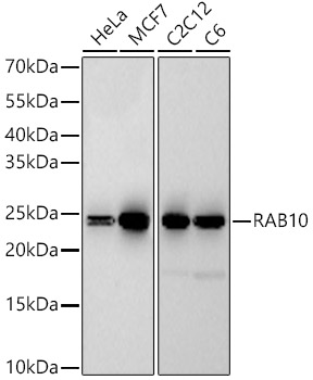 | Western blot analysis of various lysates, using [KD Validated] RAB10 Rabbit mAb (A22746) at 1:60000 dilution. Secondary antibody: HRP-conjugated Goat anti-Rabbit IgG (H+L) (AS014) at 1:10000 dilution. Lysates/proteins: 25μg per lane. Blocking buffer: 3% nonfat dry milk in TBST. Detection: ECL Basic Kit (RM00020). Exposure time: 180s. |
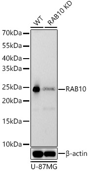 | Western blot analysis of lysates from wild type(WT) and RAB10 knockdown (KD) U-87MG cells, using [KD Validated] RAB10 Rabbit mAb (A22746) at 1:60000 dilution. Secondary antibody: HRP-conjugated Goat anti-Rabbit IgG (H+L) (AS014) at 1:10000 dilution. Lysates/proteins: 25μg per lane. Blocking buffer: 3% nonfat dry milk in TBST. Detection: ECL Basic Kit (RM00020). Exposure time: 180s. |
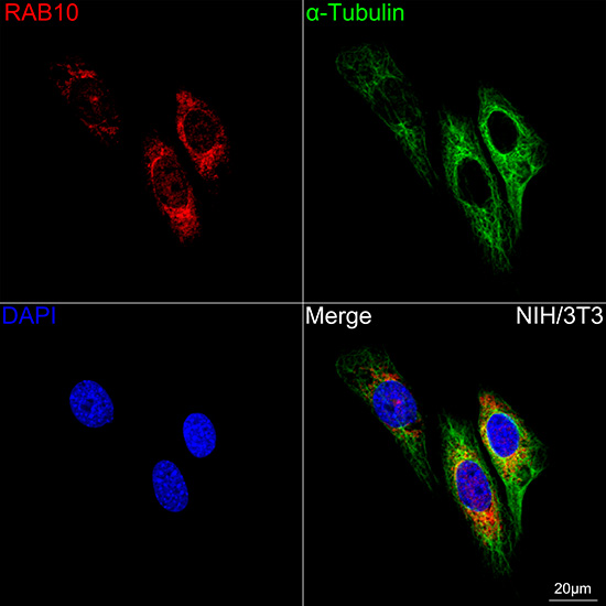 | Confocal imaging of NIH/3T3 cells using [KD Validated] RAB10 Rabbit mAb (A22746, dilution 1:200) followed by a further incubation with Cy3 Goat Anti-Rabbit IgG (H+L) (AS007, dilution 1:500) (Red). The cells were counterstained with α-Tubulin Mouse mAb (AC012, dilution 1:400) followed by incubation with ABflo® 488-conjugated Goat Anti-Mouse IgG (H+L) Ab (AS076, dilution 1:500) (Green). DAPI was used for nuclear staining (Blue). Objective: 100x. |
You may also be interested in:

