Your shopping cart is empty!
")
KD-Validated CD98/SLC3A2 Rabbit PolymAb® (20 μl)
| Reactivity: | Human, Rat |
| Applications: | WB, IF/IC, ELISA |
| Host Species: | Rabbit |
| Isotype: | IgG |
| Clonality: | Monoclonal antibody |
| Gene Name: | solute carrier family 3 member 2 |
| Gene Symbol: | SLC3A2 |
| Synonyms: | 4F2; CD98; MDU1; 4F2HC; 4T2HC; NACAE; CD98HC |
| Gene ID: | 6520/17254 |
| UniProt ID: | P08195/P10852 |
| Immunogen: | Recombinant fusion protein containing a sequence corresponding to amino acids 206-630 of human/mouse SLC3A2/CD98hc (NP_002385.3). |
| Purification Method: | Affinity purification |
| Dilution: | WB 1:1000-1:6000; IF/IC 1:200-1:800 |
| Concentration: | 1.57mg/ml |
| Buffer: | PBS with 0.09% Sodium azide, 0.05% BSA, 50% glycerol, pH7.3. |
| Storage: | Store at -20°C. Avoid freeze / thaw cycles. |
| Documents: | Manual-SLC3A2 monoclonal antibody |
Background
The gene SLC3A2 is a member of the solute carrier family and encodes a cell surface, transmembrane protein. The protein exists as the heavy chain of a heterodimer, covalently bound through di-sulfide bonds to one of several possible light chains. The encoded transporter plays a role in regulation of intracellular calcium levels and transports L-type amino acids. Alternatively spliced transcript variants, encoding different isoforms, have been characterized.
Images
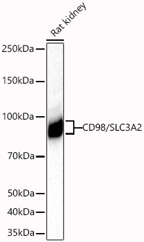 | Western blot analysis of lysates from Rat kidney using [KD Validated]CD98/SLC3A2 Rabbit PolymAb® (A19880PM) at 1:1000 dilution incubated at room temperature for 1.5 hours. Secondary antibody: HRP-conjugated Goat anti-Rabbit IgG (H+L) (AS014) at 1:10000 dilution. Lysates/proteins: 25 μg per lane. Blocking buffer: 3% nonfat dry milk in TBST. Detection: ECL Basic Kit (RM00020). Exposure time: 90s. |
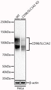 | Western blot analysis of lysates from wild type (WT) and CD98/SLC3A2 knockdown (KD) HeLa cells using [KD Validated]CD98/SLC3A2 Rabbit PolymAb® (A19880PM) at 1:1000 dilution incubated at room temperature for 1.5 hours. Secondary antibody: HRP-conjugated Goat anti-Rabbit IgG (H+L) (AS014) at 1:10000 dilution. Lysates/proteins: 25 μg per lane. Blocking buffer: 3% nonfat dry milk in TBST. Detection: ECL Basic Kit (RM00020). Exposure time: 5s. |
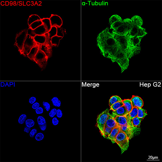 | Confocal imaging of Hep G2 cells using [KD Validated]CD98/SLC3A2 Rabbit PolymAb® (A19880PM, dilution 1:200) followed by a further incubation with Cy3 Goat Anti-Rabbit IgG (H+L) (AS007, dilution 1:500) (Red). The cells were counterstained with α-Tubulin Mouse mAb (AC012, dilution 1:400) followed by incubation with ABflo® 488-conjugated Goat Anti-Mouse IgG (H+L) Ab (AS076, dilution 1:500) (Green). DAPI was used for nuclear staining (Blue). Objective: 100x. |
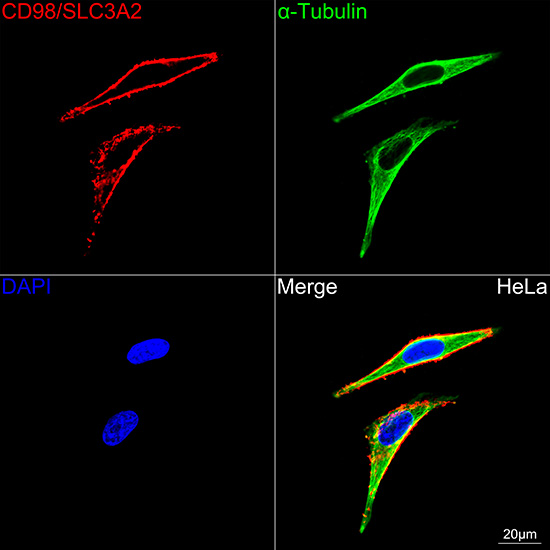 | Confocal imaging of HeLa cells using [KD Validated]CD98/SLC3A2 Rabbit PolymAb® (A19880PM, dilution 1:200) followed by a further incubation with Cy3 Goat Anti-Rabbit IgG (H+L) (AS007, dilution 1:500) (Red). The cells were counterstained with α-Tubulin Mouse mAb (AC012, dilution 1:400) followed by incubation with ABflo® 488-conjugated Goat Anti-Mouse IgG (H+L) Ab (AS076, dilution 1:500) (Green). DAPI was used for nuclear staining (Blue). Objective: 100x. |
You may also be interested in:

