Your shopping cart is empty!
")
| Reactivity: | Human, Mouse, Rat |
| Applications: | WB, IHC, IF/IC, ELISA |
| Host Species: | Rabbit |
| Isotype: | IgG |
| Clonality: | Monoclonal antibody |
| Gene Name: | signal recognition particle 72 |
| Gene Symbol: | SRP72 |
| Synonyms: | BMFF; BMFS1; HEL103; SRP72 |
| Gene ID: | 6731 |
| UniProt ID: | O76094 |
| Clone ID: | 5Q0Y8 |
| Immunogen: | Recombinant fusion protein containing a sequence corresponding to amino acids 440-544 of human SRP72 (NP_008878.3). |
| Dilution: | WB 1:3000-1:12000; IHC 1:200-1:800; IF/IC 1:200-1:400 |
| Purification Method: | Affinity purification |
| Concentration: | 1.88 mg/mL |
| Buffer: | PBS with 0.05% proclin300, 0.05% BSA, 50% glycerol, pH7.3. |
| Storage: | Store at -20°C. Avoid freeze / thaw cycles. |
| Documents: | Manual-SRP72 monoclonal antibody |
Background
This gene encodes the 72 kDa subunit of the signal recognition particle (SRP), a ribonucleoprotein complex that mediates the targeting of secretory proteins to the endoplasmic reticulum (ER). The SRP complex consists of a 7S RNA and 6 protein subunits: SRP9, SRP14, SRP19, SRP54, SRP68, and SRP72, that are bound to the 7S RNA as monomers or heterodimers. SRP has at least 3 distinct functions that can be associated with the protein subunits: signal recognition, translational arrest, and ER membrane targeting by interaction with the docking protein. Mutations in this gene are associated with familial bone marrow failure. Alternatively spliced transcript variants encoding different isoforms have been found for this gene.
Images
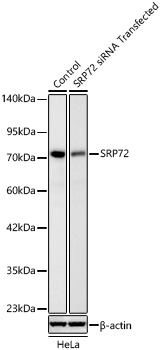 | Western blot analysis of lysates from HeLa cells transfected with SRP72 siRNA using [KD Validated] SRP72 Rabbit mAb (A25418) at 1:3000 dilution. Secondary antibody:HRP Goat Anti-Rabbit IgG (H+L) (AS014) at 1:10000 dilution. Lysates/proteins: 18 μg per lane. Blocking buffer: 3% nonfat dry milk in TBST. Detection: ECL Basic Kit (RM00020). Exposure time: 30s. |
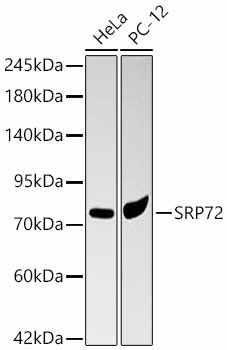 | Western blot analysis of various lysates using [KD Validated] SRP72 Rabbit mAb (A25418) at 1:3000 dilution. Secondary antibody:HRP Goat Anti-Rabbit IgG (H+L) (AS014) at 1:10000 dilution. Lysates / proteins: 25 μg per lane. Blocking buffer: 3 % nonfat dry milk in TBST. Detection: ECL Basic Kit (RM00020). Exposure time: 5s. |
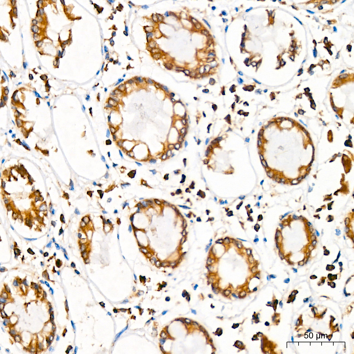 | Immunohistochemistry analysis of paraffin-embedded Human colon tissue using [KD Validated] SRP72 Rabbit mAb (A25418) at a dilution of 1:200 (40x lens). High pressure antigen retrieval was performed with 0.01 M citrate buffer (pH 6.0) prior to IHC staining. |
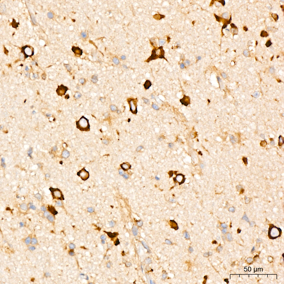 | Immunohistochemistry analysis of paraffin-embedded Human brain tissue using [KD Validated] SRP72 Rabbit mAb (A25418) at a dilution of 1:200 (40x lens). High pressure antigen retrieval was performed with 0.01 M citrate buffer (pH 6.0) prior to IHC staining. |
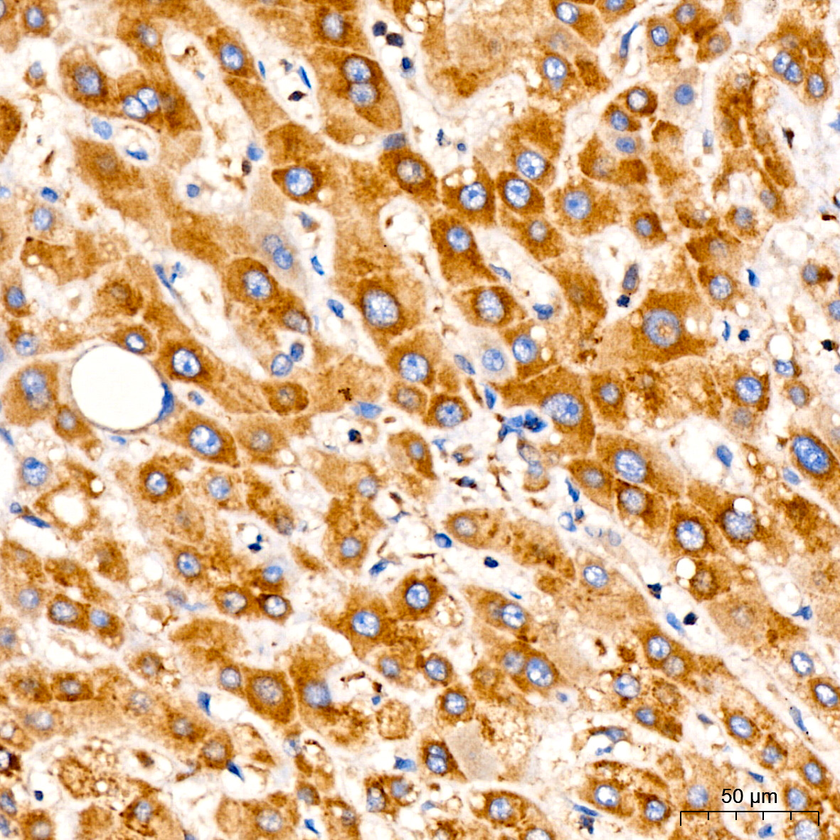 | Immunohistochemistry analysis of paraffin-embedded Human liver tissue using [KD Validated] SRP72 Rabbit mAb (A25418) at a dilution of 1:200 (40x lens). High pressure antigen retrieval was performed with 0.01 M citrate buffer (pH 6.0) prior to IHC staining. |
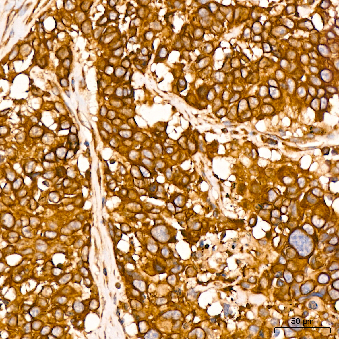 | Immunohistochemistry analysis of paraffin-embedded Human lung cancer tissue using [KD Validated] SRP72 Rabbit mAb (A25418) at a dilution of 1:200 (40x lens). High pressure antigen retrieval was performed with 0.01 M citrate buffer (pH 6.0) prior to IHC staining. |
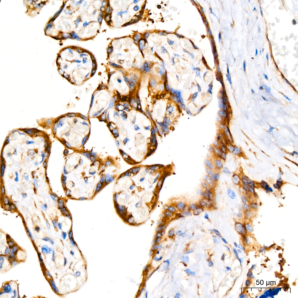 | Immunohistochemistry analysis of paraffin-embedded Human placenta tissue using [KD Validated] SRP72 Rabbit mAb (A25418) at a dilution of 1:200 (40x lens). High pressure antigen retrieval was performed with 0.01 M citrate buffer (pH 6.0) prior to IHC staining. |
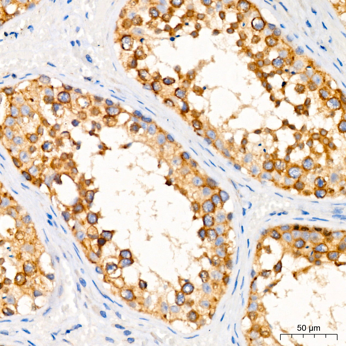 | Immunohistochemistry analysis of paraffin-embedded Human testis tissue using [KD Validated] SRP72 Rabbit mAb (A25418) at a dilution of 1:200 (40x lens). High pressure antigen retrieval was performed with 0.01 M citrate buffer (pH 6.0) prior to IHC staining. |
You may also be interested in:

