Your shopping cart is empty!
")
| Reactivity: | Human, Mouse, Rat |
| Applications: | WB, ELISA |
| Host Species: | Rabbit |
| Isotype: | IgG |
| Clonality: | Polyclonal antibody |
| Gene Name: | voltage dependent anion channel 1 |
| Gene Symbol: | VDAC1 |
| Synonyms: | PORIN; VDAC-1; [KD Validated] VDAC1 |
| Gene ID: | 7416 |
| UniProt ID: | P21796 |
| Immunogen: | Recombinant fusion protein containing a sequence corresponding to amino acids 1-283 of human VDAC1 (NP_003365.1). |
| Dilution: | WB 1:500-1:1000 |
| Purification Method: | Affinity purification |
| Concentration: | 0.55 mg/ml |
| Buffer: | PBS with 0.05% proclin300, 50% glycerol, pH7.3. |
| Storage: | Store at -20°C. Avoid freeze / thaw cycles. |
| Documents: | Manual-VDAC1 polyclonal antibody |
Background
This gene encodes a voltage-dependent anion channel protein that is a major component of the outer mitochondrial membrane. The encoded protein facilitates the exchange of metabolites and ions across the outer mitochondrial membrane and may regulate mitochondrial functions. This protein also forms channels in the plasma membrane and may be involved in transmembrane electron transport. Alternate splicing results in multiple transcript variants. Multiple pseudogenes of this gene are found on chromosomes 1, 2 3, 6, 9, 12, X and Y.
Images
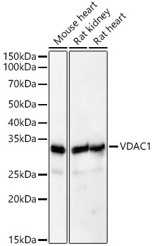 | Western blot analysis of various lysates, using [KD Validated] VDAC1 Rabbit pAb (A21730) at 1:600 dilution. Secondary antibody: HRP-conjugated Goat anti-Rabbit IgG (H+L) (AS014) at 1:10000 dilution. Lysates/proteins: 25μg per lane. Blocking buffer: 3% nonfat dry milk in TBST. Detection: ECL Basic Kit (RM00020). Exposure time: 10s. |
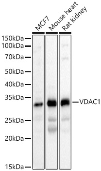 | Western blot analysis of various lysates, using [KD Validated] VDAC1 Rabbit pAb (A21730) at 1:500 dilution. Secondary antibody: HRP-conjugated Goat anti-Rabbit IgG (H+L) (AS014) at 1:10000 dilution. Lysates/proteins: 25μg per lane. Blocking buffer: 3% nonfat dry milk in TBST. Detection: ECL Basic Kit (RM00020). Exposure time: 10s. |
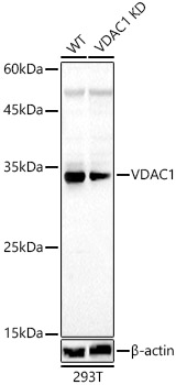 | Western blot analysis of lysates from wild type (WT) and VDAC1 knockdown (KD) 293T cells, using [KD Validated] VDAC1 Rabbit pAb (A21730) at 1:1000 dilution. Secondary antibody: HRP-conjugated Goat anti-Rabbit IgG (H+L) (AS014) at 1:10000 dilution. Lysates/proteins: 25μg per lane. Blocking buffer: 3% nonfat dry milk in TBST. Detection: ECL Basic Kit (RM00020). Exposure time: 60s. |
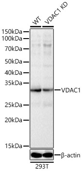 | Western blot analysis of lysates from wild type(WT) and VDAC1 knockdown (KD) 293T cells, using [KD Validated] VDAC1 Rabbit pAb (A21730) at 1:600 dilution. Secondary antibody: HRP-conjugated Goat anti-Rabbit IgG (H+L) (AS014) at 1:10000 dilution. Lysates/proteins: 25μg per lane. Blocking buffer: 3% nonfat dry milk in TBST. Detection: ECL Basic Kit (RM00020). Exposure time: 10s. |
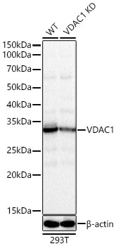 | Western blot analysis of lysates from wild type(WT) and VDAC1 knockdown (KD) 293T cells, using [KD Validated] VDAC1 Rabbit pAb (A21730) at 1:500 dilution. Secondary antibody: HRP-conjugated Goat anti-Rabbit IgG (H+L) (AS014) at 1:10000 dilution. Lysates/proteins: 25μg per lane. Blocking buffer: 3% nonfat dry milk in TBST. Detection: ECL Basic Kit (RM00020). Exposure time: 10s. |
You may also be interested in:

