Your shopping cart is empty!
| Reactivity: | Human |
| Applications: | WB, ELISA |
| Host Species: | Rabbit |
| Isotype: | IgG |
| Clonality: | Polyclonal antibody |
| Gene Name: | autophagy related 4A cysteine peptidase |
| Gene Symbol: | ATG4A |
| Synonyms: | APG4A; AUTL2; HsAPG4A; ATG4A |
| Gene ID: | 115201 |
| UniProt ID: | Q8WYN0 |
| Immunogen: | Recombinant fusion protein containing a sequence corresponding to amino acids 289-398 of human ATG4A (NP_443168.2). |
| Purification Method: | Affinity purification |
| Concentration: | 0.85 mg/mL |
| Buffer: | PBS with 0.02% sodium azide, 50% glycerol ,pH7.3. |
| Storage: | Store at -20°C. Avoid freeze / thaw cycles. |
| Documents: | Manual-ATG4A polyclonal antibody |
Background
Autophagy is the process by which endogenous proteins and damaged organelles are destroyed intracellularly. Autophagy is postulated to be essential for cell homeostasis and cell remodeling during differentiation, metamorphosis, non-apoptotic cell death, and aging. Reduced levels of autophagy have been described in some malignant tumors, and a role for autophagy in controlling the unregulated cell growth linked to cancer has been proposed. This gene encodes a member of the autophagin protein family. The encoded protein is also designated as a member of the C-54 family of cysteine proteases.
Images
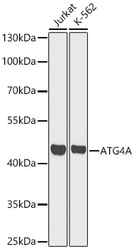 | Western blot analysis of various lysates using [KO Validated] ATG4A Rabbit pAb (A21356) at 1:1000 dilution. Secondary antibody: HRP-conjugated Goat anti-Rabbit IgG (H+L) (AS014) at 1:10000 dilution. Lysates / proteins: 25 μg per lane. Blocking buffer: 3 % nonfat dry milk in TBST. Detection: ECL Basic Kit (RM00020). Exposure time: 30s. |
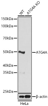 | Western blot analysis of lysates from wild type (WT) and ATG4A knockout (KO) HeLa cells using [KO Validated] ATG4A Rabbit pAb (A21356) at 1:1000 dilution. Secondary antibody: HRP-conjugated Goat anti-Rabbit IgG (H+L) (AS014) at 1:10000 dilution.Lysates/proteins: 25 μg per lane. Blocking buffer: 3% nonfat dry milk in TBST. Detection: ECL Basic Kit (RM00020). Exposure time: 5s. |
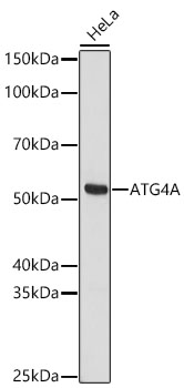 | Western blot analysis of lysates from HeLa cells using [KO Validated] ATG4A Rabbit pAb (A21356) at 1:1000 dilution. Secondary antibody: HRP-conjugated Goat anti-Rabbit IgG (H+L) (AS014) at 1:10000 dilution. Lysates / proteins: 25 μg per lane. Blocking buffer: 3 % nonfat dry milk in TBST. Detection: ECL Basic Kit (RM00020). Exposure time: 30s. |
You may also be interested in:

