Your shopping cart is empty!
KO-Validated CHCHD2 Rabbit pAb (20 μl)
| Reactivity: | Human |
| Applications: | WB, IHC-P, IF/ICC, ELISA |
| Host Species: | Rabbit |
| Isotype: | IgG |
| Clonality: | Polyclonal antibody |
| Gene Name: | coiled-coil-helix-coiled-coil-helix domain containing 2 |
| Gene Symbol: | CHCHD2 |
| Synonyms: | MNRR1; NS2TP; MIX17B; PARK22; C7orf17; D2 |
| Gene ID: | 51142 |
| UniProt ID: | Q9Y6H1 |
| Immunogen: | Recombinant fusion protein containing a sequence corresponding to amino acids 75-145 of human CHCHD2 (NP_057223.1). |
| Dilution: | WB 1:500-1:2000; IF/IC 1:50-1:200 |
| Purification Method: | Affinity purification |
| Concentration: | 0.59 mg/mL |
| Buffer: | PBS with 0.01% thimerosal, 50% glycerol, pH7.3. |
| Storage: | Store at -20°C. Avoid freeze / thaw cycles. |
| Documents: | Manual-CHCHD2 polyclonal antibody |
Background
The protein encoded by this gene belongs to a class of eukaryotic CX(9)C proteins characterized by four cysteine residues spaced ten amino acids apart from one another. These residues form disulfide linkages that define a CHCH fold. In response to stress, the protein translocates from the mitochondrial intermembrane space to the nucleus where it binds to a highly conserved 13 nucleotide oxygen responsive element in the promoter of cytochrome oxidase 4I2, a subunit of the terminal enzyme of the electron transport chain. In concert with recombination signal sequence-binding protein J, binding of this protein activates the oxygen responsive element at four percent oxygen. In addition, it has been shown that this protein is a negative regulator of mitochondria-mediated apoptosis. In response to apoptotic stimuli, mitochondrial levels of this protein decrease, allowing BCL2-associated X protein to oligomerize and activate the caspase cascade. Pseudogenes of this gene are found on multiple chromosomes. Alternative splicing results in multiple transcript variants.
Images
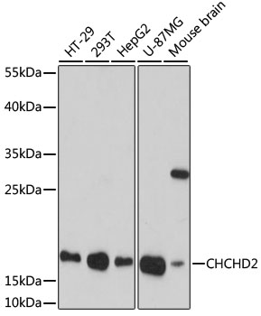 | Western blot analysis of various lysates using [KO Validated] CHCHD2 Rabbit pAb (A16645) at 1:1000 dilution. Secondary antibody: HRP-conjugated Goat anti-Rabbit IgG (H+L) (AS014) at 1:10000 dilution. Lysates/proteins: 25μg per lane. Blocking buffer: 3% nonfat dry milk in TBST. Detection: ECL Basic Kit (RM00020). Exposure time: 90s. |
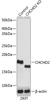 | Western blot analysis of lysates from wild type (WT) and CHCHD2 knockout (KO) 293T cells, using [KO Validated] CHCHD2 Rabbit pAb (A16645) at 1:1000 dilution. Secondary antibody: HRP-conjugated Goat anti-Rabbit IgG (H+L) (AS014) at 1:10000 dilution. Lysates/proteins: 25μg per lane. Blocking buffer: 3% nonfat dry milk in TBST. Detection: ECL Basic Kit (RM00020). Exposure time: 1s. |
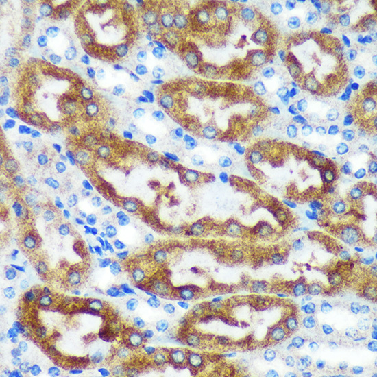 | Immunohistochemistry analysis of paraffin-embedded Mouse kidney using [KO Validated] CHCHD2 Rabbit pAb (A16645) at dilution of 1:100 (40x lens). Microwave antigen retrieval performed with 0.01M PBS Buffer (pH 7.2) prior to IHC staining. |
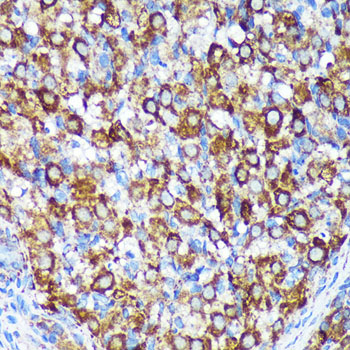 | Immunohistochemistry analysis of paraffin-embedded Rat ovary using [KO Validated] CHCHD2 Rabbit pAb (A16645) at dilution of 1:100 (40x lens). Microwave antigen retrieval performed with 0.01M PBS Buffer (pH 7.2) prior to IHC staining. |
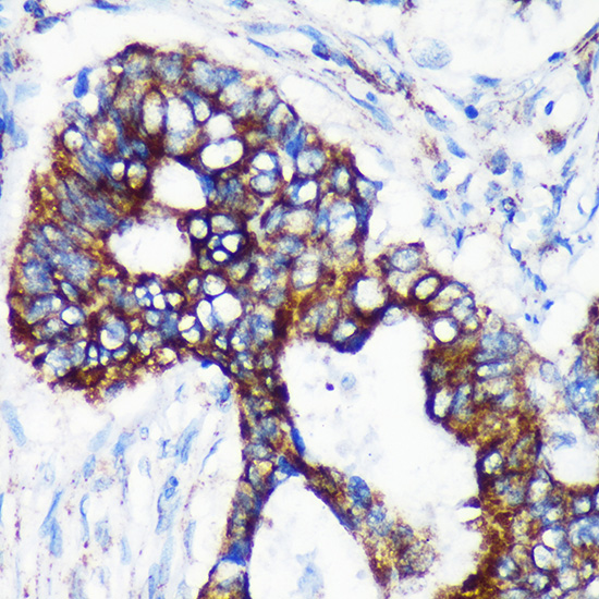 | Immunohistochemistry analysis of paraffin-embedded Human colon carcinoma using [KO Validated] CHCHD2 Rabbit pAb (A16645) at dilution of 1:100 (40x lens). Microwave antigen retrieval performed with 0.01M PBS Buffer (pH 7.2) prior to IHC staining. |
 | Immunofluorescence analysis of C6 cells using [KO Validated] CHCHD2 Rabbit pAb (A16645) at dilution of 1:100. Secondary antibody: Cy3-conjugated Goat anti-Rabbit IgG (H+L) (AS007) at 1:500 dilution. Blue: DAPI for nuclear staining. |
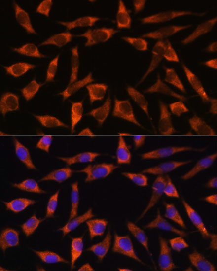 | Immunofluorescence analysis of L929 cells using [KO Validated] CHCHD2 Rabbit pAb (A16645) at dilution of 1:100. Secondary antibody: Cy3-conjugated Goat anti-Rabbit IgG (H+L) (AS007) at 1:500 dilution. Blue: DAPI for nuclear staining. |
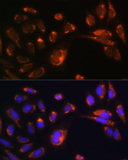 | Immunofluorescence analysis of U-2 OS cells using [KO Validated] CHCHD2 Rabbit pAb (A16645) at dilution of 1:100. Secondary antibody: Cy3-conjugated Goat anti-Rabbit IgG (H+L) (AS007) at 1:500 dilution. Blue: DAPI for nuclear staining. |
You may also be interested in:

