Your shopping cart is empty!
")
| Reactivity: | Human,Mouse |
| Applications: | WB, IHC, ELISA |
| Host Species: | Rabbit |
| Isotype: | IgG |
| Clonality: | Monoclonal antibody |
| Gene Name: | mutL homolog 1 |
| Gene Symbol: | MLH1 |
| Synonyms: | FCC2; COCA2; HNPCC; MLH-1; hMLH1; HNPCC2; LYNCH2; MMRCS1 |
| Gene ID: | 4292 |
| UniProt ID: | P40692 |
| Clone ID: | 8K1T2 |
| Immunogen: | A synthetic peptide corresponding to a sequence within amino acids 350-450 of human MLH1 (NP_000240.1). |
| Dilution: | WB 1:10000-1:70000; IHC 1:100-1:500 |
| Purification Method: | Affinity purification |
| Concentration: | 0.69 mg/mL |
| Buffer: | PBS with 0.05% proclin300, 0.05% BSA, 50% glycerol, pH7.3. |
| Storage: | Store at -20°C. Avoid freeze / thaw cycles. |
| Documents: | Manual-MLH1 monoclonal antibody |
Background
The protein encoded by this gene can heterodimerize with mismatch repair endonuclease PMS2 to form MutL alpha, part of the DNA mismatch repair system. When MutL alpha is bound by MutS beta and some accessory proteins, the PMS2 subunit of MutL alpha introduces a single-strand break near DNA mismatches, providing an entry point for exonuclease degradation. The encoded protein is also involved in DNA damage signaling and can heterodimerize with DNA mismatch repair protein MLH3 to form MutL gamma, which is involved in meiosis. This gene was identified as a locus frequently mutated in hereditary nonpolyposis colon cancer (HNPCC).
Images
 | Western blot analysis of lysates from mouse brain, using [KO Validated] MLH1 Rabbit mAb (A4858) at1:60000 dilution. Secondary antibody: HRP-conjugated Goat anti-Rabbit IgG (H+L) (AS014) at 1:10000 dilution. Lysates/proteins: 25μg per lane. Blocking buffer: 3% nonfat dry milk in TBST. Detection: ECL Basic Kit (RM00020). Exposure time: 180s. |
 | Western blot analysis of lysates from wild type (WT) and MLH1 knockout (KO) HeLa cells, using [KO Validated] MLH1 Rabbit mAb (A4858) at1:60000 dilution. Secondary antibody: HRP-conjugated Goat anti-Rabbit IgG (H+L) (AS014) at 1:10000 dilution. Lysates/proteins: 25μg per lane. Blocking buffer: 3% nonfat dry milk in TBST. Detection: ECL Basic Kit (RM00020). Exposure time: 180s. |
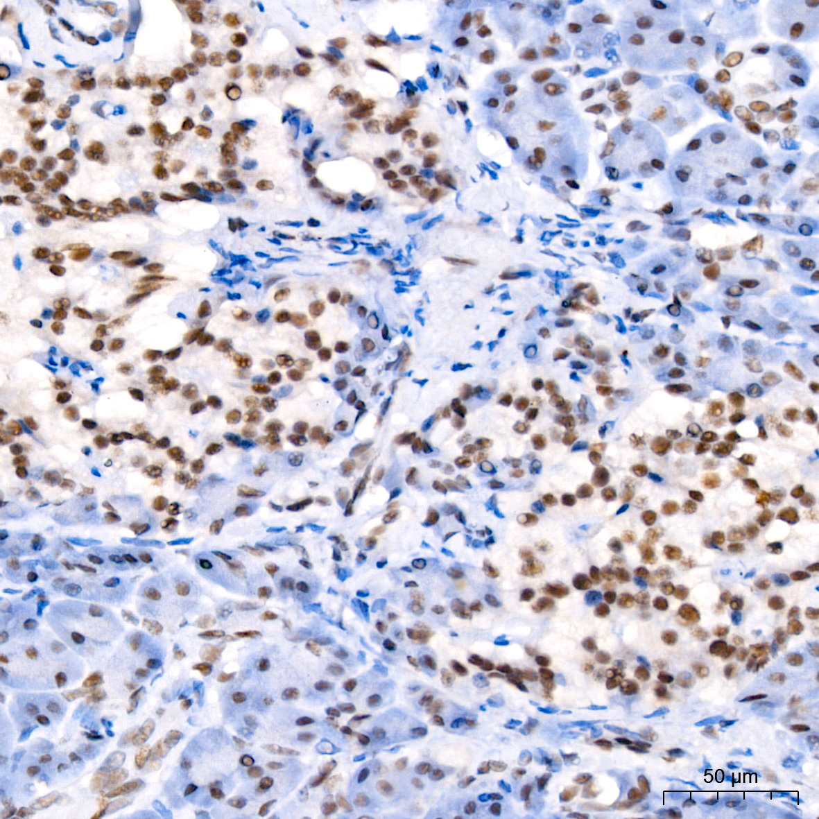
 | Immunohistochemistry analysis of paraffin-embedded Human colon carcinoma using [KO Validated] MLH1 Rabbit mAb (A4858) at dilution of 1:100 (40x lens). High pressure antigen retrieval performed with 0.01M Citrate Bufferr (pH 6.0) prior to IHC staining. |
 | Immunohistochemistry analysis of paraffin-embedded Human colon carcinoma tissue using [KO Validated] MLH1 Rabbit mAb (A25641) at a dilution of 1:100 (40x lens). High pressure antigen retrieval performed with 0.01M Citrate Buffer (pH 6.0) prior to IHC staining. |
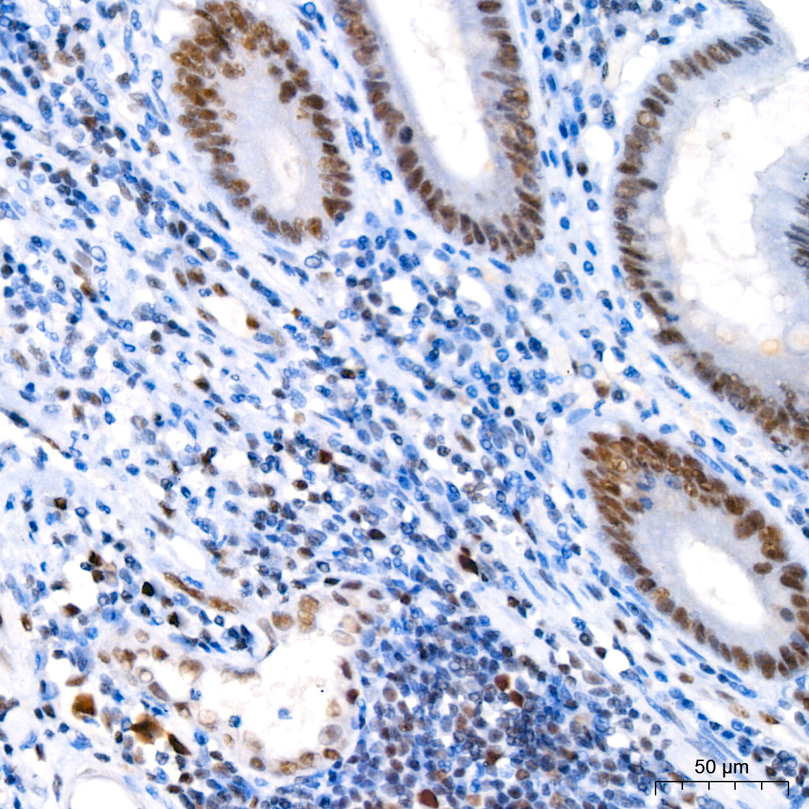 | Immunohistochemistry analysis of paraffin-embedded Human appendix tissue using [KO Validated] MLH1 Rabbit mAb (A25641) at a dilution of 1:100 (40x lens). High pressure antigen retrieval performed with 0.01M Citrate Buffer (pH 6.0) prior to IHC staining. |
 | Immunohistochemistry analysis of paraffin-embedded Human pancreas tissue using [KO Validated] MLH1 Rabbit mAb (A25641) at a dilution of 1:100 (40x lens). High pressure antigen retrieval performed with 0.01M Citrate Buffer (pH 6.0) prior to IHC staining. |
 | Immunohistochemistry analysis of paraffin-embedded Human tonsil tissue using [KO Validated] MLH1 Rabbit mAb (A25641) at a dilution of 1:100 (40x lens). High pressure antigen retrieval performed with 0.01M Citrate Buffer (pH 6.0) prior to IHC staining. |
You may also be interested in:

