Your shopping cart is empty!
")
| Reactivity: | Human |
| Applications: | WB, ELISA, ChIP, CUT&Tag |
| Host Species: | Rabbit |
| Isotype: | IgG |
| Clonality: | Monoclonal antibody |
| Gene Name: | MYC proto-oncogene, bHLH transcription factor |
| Gene Symbol: | MYC |
| Synonyms: | MRTL; MYCC; c-Myc; bHLHe39; yc |
| Gene ID: | 4609 |
| UniProt ID: | P01106 |
| Immunogen: | A synthetic peptide corresponding to a sequence within amino acids 1-100 of human c-Myc (P01106). |
| Dilution: | WB 1:1000-1:2000 |
| Purification Method: | Affinity purification |
| Buffer: | PBS with 0.02% sodium azide, 0.05% BSA, 50% glycerol, pH7.3. |
| Storage: | Store at -20°C. Avoid freeze / thaw cycles. |
| Documents: | Manual-MYC monoclonal antibody |
Background
This gene is a proto-oncogene and encodes a nuclear phosphoprotein that plays a role in cell cycle progression, apoptosis and cellular transformation. The encoded protein forms a heterodimer with the related transcription factor MAX. This complex binds to the E box DNA consensus sequence and regulates the transcription of specific target genes. Amplification of this gene is frequently observed in numerous human cancers. Translocations involving this gene are associated with Burkitt lymphoma and multiple myeloma in human patients. There is evidence to show that translation initiates both from an upstream, in-frame non-AUG (CUG) and a downstream AUG start site, resulting in the production of two isoforms with distinct N-termini.
Images
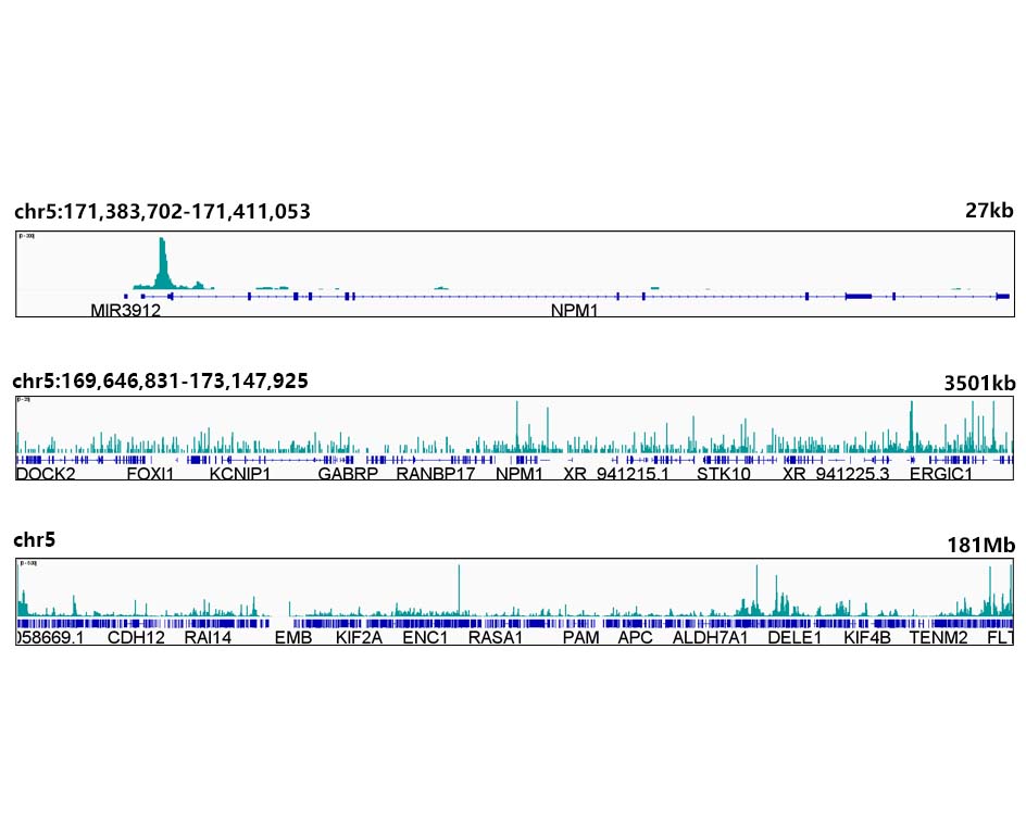 | UT&Tag was performed using the CUT&Tag Assay Kit (pAG-Tn5) for Illumina(RK20265) from 10⁵ K562 cells with 1μg c-Myc Rabbit mAb(A19032), along with a Goat Anti-Rabbit IgG(H+L). The CUT&Tag results indicate the enrichment pattern of ESE1 in representative gene loci (NPM1), as shown in figure. |
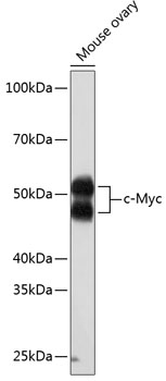
 | Western blot analysis of various lysates using [KO Validated] c-Myc Rabbit mAb (A19032) at 1:1000 dilution. Secondary antibody: HRP-conjugated Goat anti-Rabbit IgG (H+L) (AS014) at 1:10000 dilution. Lysates/proteins: 25μg per lane. Blocking buffer: 3% nonfat dry milk in TBST. Detection: ECL Basic Kit (RM00020). Exposure time: 10s. |
 | Western blot analysis of lysates from wild type (WT) and c-Myc knockout (KO) HCT 116 cells, using [KO Validated] c-Myc Rabbit mAb (A19032) at 1:1000 dilution. Secondary antibody: HRP-conjugated Goat anti-Rabbit IgG (H+L) (AS014) at 1:10000 dilution. Lysates/proteins: 25μg per lane. Blocking buffer: 3% nonfat dry milk in TBST. Detection: ECL Basic Kit (RM00020). Exposure time: 10s. |
 | Chromatin immunoprecipitation analysis of extracts of K562 cells, using c-Myc antibody (A19032) and rabbit IgG.The amount of immunoprecipitated DNA was checked by quantitative PCR. Histogram was constructed by the ratios of the immunoprecipitated DNA to the input. |
You may also be interested in:

