| Reactivity: | Human |
| Applications: | WB, IHC-P, IF/ICC, ELISA |
| Host Species: | Rabbit |
| Isotype: | IgG |
| Clonality: | Polyclonal antibody |
| Gene Name: | proteasome activator subunit 3 |
| Gene Symbol: | PSME3 |
| Synonyms: | Ki; PA28G; HEL-S-283; PA28gamma; REG-GAMMA; PA28-gamma; E3 |
| Gene ID: | 10197 |
| UniProt ID: | P61289 |
| Immunogen: | A synthetic peptide corresponding to a sequence within amino acids 200-254 of human PSME3 (NP_005780.2). |
| Dilution: | WB 1:100-1:500; IHC 1:50-1:200 |
| Purification Method: | Affinity purification |
| Concentration: | 0.98 mg/ml |
| Buffer: | PBS with 0.01% thimerosal, 50% glycerol, pH7.3. |
| Storage: | Store at -20°C. Avoid freeze / thaw cycles. |
| Documents: | Manual-PSME3 polyclonal antibody |
Background
The 26S proteasome is a multicatalytic proteinase complex with a highly ordered structure composed of 2 complexes, a 20S core and a 19S regulator. The 20S core is composed of 4 rings of 28 non-identical subunits; 2 rings are composed of 7 alpha subunits and 2 rings are composed of 7 beta subunits. The 19S regulator is composed of a base, which contains 6 ATPase subunits and 2 non-ATPase subunits, and a lid, which contains up to 10 non-ATPase subunits. Proteasomes are distributed throughout eukaryotic cells at a high concentration and cleave peptides in an ATP/ubiquitin-dependent process in a non-lysosomal pathway. An essential function of a modified proteasome, the immunoproteasome, is the processing of class I MHC peptides. The immunoproteasome contains an alternate regulator, referred to as the 11S regulator or PA28, that replaces the 19S regulator. Three subunits (alpha, beta and gamma) of the 11S regulator have been identified. This gene encodes the gamma subunit of the 11S regulator. Six gamma subunits combine to form a homohexameric ring. Alternate splicing results in multiple transcript variants.
Images
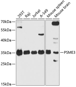 | Western blot analysis of various lysates using PSME3 Rabbit pAb (A18021) at 1:3000 dilution. Secondary antibody: HRP-conjugated Goat anti-Rabbit IgG (H+L) (AS014) at 1:10000 dilution. Lysates/proteins: 25μg per lane. Blocking buffer: 3% nonfat dry milk in TBST. Detection: ECL Basic Kit (RM00020). Exposure time: 30s. |
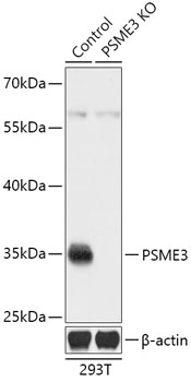 | Western blot analysis of lysates from wild type (WT) and PSME3 knockout (KO) 293T cells, using [KO Validated] PSME3 Rabbit pAb (A18021) at 1:3000 dilution. Secondary antibody: HRP-conjugated Goat anti-Rabbit IgG (H+L) (AS014) at 1:10000 dilution. Lysates/proteins: 25μg per lane. Blocking buffer: 3% nonfat dry milk in TBST. Detection: ECL Basic Kit (RM00020). Exposure time: 3min. |
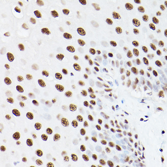 | Immunohistochemistry analysis of paraffin-embedded Human esophageal using PSME3 Rabbit pAb (A18021) at dilution of 1:100 (40x lens). Microwave antigen retrieval performed with 0.01M PBS Buffer (pH 7.2) prior to IHC staining. |
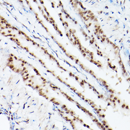 | Immunohistochemistry analysis of paraffin-embedded Mouse lung using PSME3 Rabbit pAb (A18021) at dilution of 1:100 (40x lens). Microwave antigen retrieval performed with 0.01M PBS Buffer (pH 7.2) prior to IHC staining. |
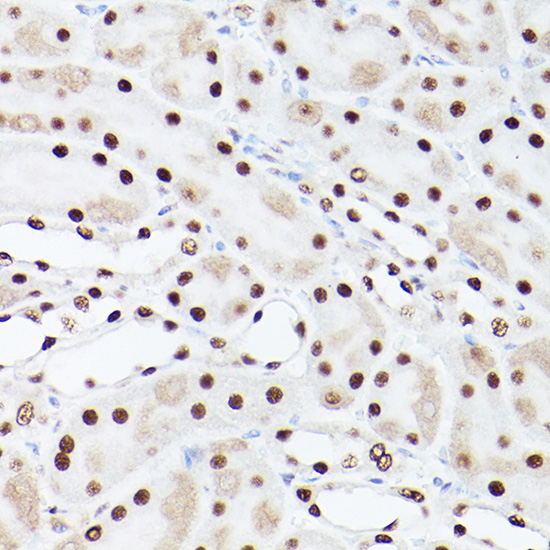 | Immunohistochemistry analysis of paraffin-embedded Rat kidney using PSME3 Rabbit pAb (A18021) at dilution of 1:100 (40x lens). Microwave antigen retrieval performed with 0.01M PBS Buffer (pH 7.2) prior to IHC staining. |
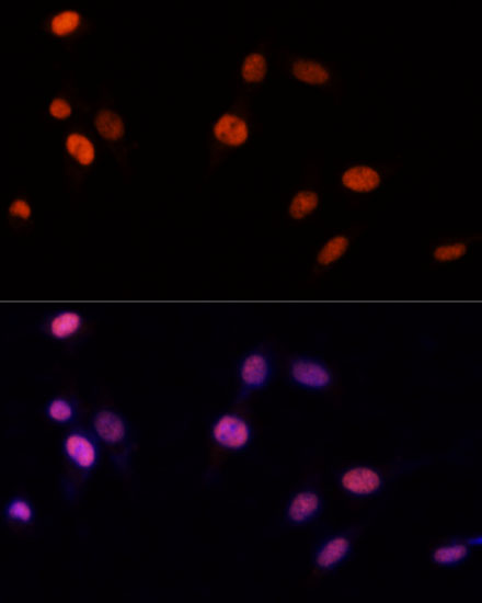 | Immunofluorescence analysis of NIH-3T3 cells using PSME3 Rabbit pAb (A18021) at dilution of 1:100 (40x lens). Secondary antibody: Cy3-conjugated Goat anti-Rabbit IgG (H+L) (AS007) at 1:500 dilution. Blue: DAPI for nuclear staining. |
You may also be interested in:

