Your shopping cart is empty!
")
| Reactivity: | Human, Mouse, Rat |
| Applications: | WB, IF/IC, IP, ELISA |
| Host Species: | Rabbit |
| Isotype: | IgG |
| Clonality: | Polyclonal antibody |
| Gene Name: | protein tyrosine phosphatase non-receptor type 11 |
| Gene Symbol: | PTPN11 |
| Synonyms: | CFC; NS1; JMML; SHP2; BPTP3; PTP2C; METCDS; PTP-1D; SH-PTP2; SH-PTP3; P2 |
| Gene ID: | 5781 |
| UniProt ID: | Q06124 |
| Immunogen: | Recombinant fusion protein containing a sequence corresponding to amino acids 1-460 of human SHP2 (NP_002825.3). |
| Dilution: | WB 1:500-1:2000; IF/IC 1:50-1:200 |
| Purification Method: | Affinity purification |
| Concentration: | 0.83 mg/ml |
| Buffer: | PBS with 0.09% Sodium azide, 50% glycerol, pH7.3. |
| Storage: | Store at -20°C. Avoid freeze / thaw cycles. |
| Documents: | Manual-PTPN11 polyclonal antibody |
Background
The protein encoded by this gene is a member of the protein tyrosine phosphatase (PTP) family. PTPs are known to be signaling molecules that regulate a variety of cellular processes including cell growth, differentiation, mitotic cycle, and oncogenic transformation. This PTP contains two tandem Src homology-2 domains, which function as phospho-tyrosine binding domains and mediate the interaction of this PTP with its substrates. This PTP is widely expressed in most tissues and plays a regulatory role in various cell signaling events that are important for a diversity of cell functions, such as mitogenic activation, metabolic control, transcription regulation, and cell migration. Mutations in this gene are a cause of Noonan syndrome as well as acute myeloid leukemia.
Images
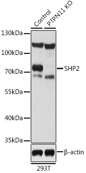 | Western blot analysis of lysates from wild type (WT) and SHP2 knockout (KO) 293T cells, using [KO Validated] SHP2 Rabbit pAb (A12486) at 1:1000 dilution. Secondary antibody: HRP-conjugated Goat anti-Rabbit IgG (H+L) (AS014) at 1:10000 dilution. Lysates/proteins: 25μg per lane. Blocking buffer: 3% nonfat dry milk in TBST. Detection: ECL Basic Kit (RM00020). Exposure time: 30s. |
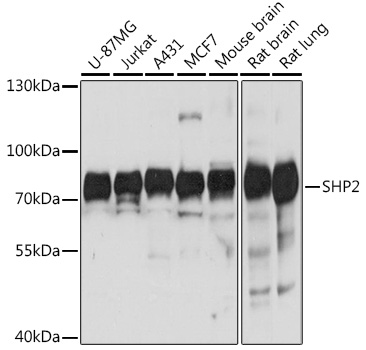 | Western blot analysis of various lysates using [KO Validated] SHP2 Rabbit pAb (A12486) at 1:1000 dilution. Secondary antibody: HRP-conjugated Goat anti-Rabbit IgG (H+L) (AS014) at 1:10000 dilution. Lysates/proteins: 25μg per lane. Blocking buffer: 3% nonfat dry milk in TBST. Detection: ECL Basic Kit (RM00020). Exposure time: 3s. |
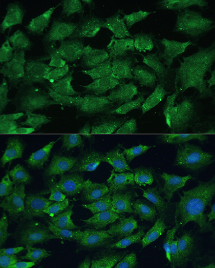 | Immunofluorescence analysis of C6 cells using [KO Validated] SHP2 Rabbit pAb (A12486) at dilution of 1:100. Blue: DAPI for nuclear staining. |
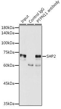 | Immunoprecipitation analysis of 300 μg extracts of MCF7 cells using 3 μg SHP2 antibody (A12486). Western blot was performed from the immunoprecipitate using SHP2 antibody (A12486) at a dilution of 1:2000. |
You may also be interested in:

