Your shopping cart is empty!
")
| Reactivity: | Human, Mouse, Rat |
| Applications: | WB, IHC, ELISA |
| Host Species: | Rabbit |
| Isotype: | IgG |
| Clonality: | Polyclonal antibody |
| Gene Name: | ribosomal protein L22 |
| Gene Symbol: | RPL22 |
| Synonyms: | EAP; L22; HBP15; HBP15/L22; 22 |
| Gene ID: | 6146 |
| UniProt ID: | P35268 |
| Immunogen: | Recombinant fusion protein containing a sequence corresponding to amino acids 1-122 of human RPL22 (NP_000974.1). |
| Dilution: | WB 1:1000-1:5000; IHC 1:50-1:100 |
| Purification Method: | Affinity purification |
| Concentration: | 0.58 mg/ml |
| Buffer: | PBS with 0.01% thimerosal, 50% glycerol, pH7.3. |
| Storage: | Store at -20°C. Avoid freeze / thaw cycles. |
| Documents: | Manual-RPL22 polyclonal antibody |
Background
Ribosomes, the organelles that catalyze protein synthesis, consist of a small 40S subunit and a large 60S subunit. Together these subunits are composed of 4 RNA species and approximately 80 structurally distinct proteins. This gene encodes a cytoplasmic ribosomal protein that is a component of the 60S subunit. The protein belongs to the L22E family of ribosomal proteins. Its initiating methionine residue is post-translationally removed. The protein can bind specifically to Epstein-Barr virus-encoded RNAs (EBERs) 1 and 2. The mouse protein has been shown to be capable of binding to heparin. Transcript variants utilizing alternative polyA signals exist. As is typical for genes encoding ribosomal proteins, there are multiple processed pseudogenes of this gene dispersed through the genome. It was previously thought that this gene mapped to 3q26 and that it was fused to the acute myeloid leukemia 1 (AML1) gene located at 21q22 in some therapy-related myelodysplastic syndrome patients with 3;21 translocations; however, these fusions actually involve a ribosomal protein L22 pseudogene located at 3q26, and this gene actually maps to 1p36.3-p36.2.
Images
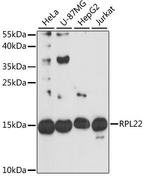 | Western blot analysis of various lysates using [KO Validated] RPL22 Rabbit pAb (A9202) at 1:3000 dilution. Secondary antibody: HRP-conjugated Goat anti-Rabbit IgG (H+L) (AS014) at 1:10000 dilution. Lysates/proteins: 25μg per lane. Blocking buffer: 3% nonfat dry milk in TBST. Detection: ECL Basic Kit (RM00020). Exposure time: 30s. |
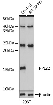 | Western blot analysis of lysates from wild type (WT) and RPL22 knockout (KO) 293T cells, using [KO Validated] RPL22 Rabbit pAb (A9202) at 1:3000 dilution. Secondary antibody: HRP-conjugated Goat anti-Rabbit IgG (H+L) (AS014) at 1:10000 dilution. Lysates/proteins: 25μg per lane. Blocking buffer: 3% nonfat dry milk in TBST. Detection: ECL Basic Kit (RM00020). Exposure time: 180s. |
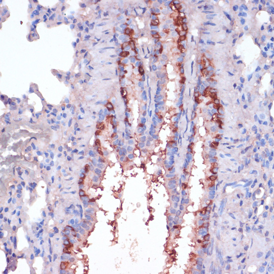 | Immunohistochemistry analysis of paraffin-embedded Rat lung using [KO Validated] RPL22 Rabbit pAb (A9202) at dilution of 1:100 (40x lens). Microwave antigen retrieval performed with 0.01M PBS Buffer (pH 7.2) prior to IHC staining. |
You may also be interested in:

