Your shopping cart is empty!
")
| Reactivity: | Human, Mouse, Rat |
| Applications: | WB, IHC, IF/IC, ELISA |
| Host Species: | Rabbit |
| Isotype: | IgG |
| Clonality: | Polyclonal antibody |
| Gene Name: | annexin A1 |
| Gene Symbol: | ANXA1 |
| Synonyms: | ANX1; LPC1; A1 |
| Gene ID: | 301 |
| UniProt ID: | P04083 |
| Immunogen: | Recombinant fusion protein containing a sequence corresponding to amino acids 1-346 of human ANXA1 (NP_000691.1). |
| Dilution: | WB 1:500-1:1000; IHC 1:50-1:200; IF/IC 1:50-1:200 |
| Purification Method: | Affinity purification |
| Concentration: | 0.92 mg/ml |
| Buffer: | PBS with 0.02% sodium azide, 50% glycerol ,pH7.3. |
| Storage: | Store at -20°C. Avoid freeze / thaw cycles. |
| Documents: | Manual-ANXA1 polyclonal antibody |
Background
The gene ANXA1 encodes a membrane-localized protein that binds phospholipids. This protein inhibits phospholipase A2 and has anti-inflammatory activity. Loss of function or expression of this gene has been detected in multiple tumors.
Images
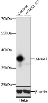 | Western blot analysis of lysates from wild type (WT) and ANXA1 knockout (KO) HeLa cells, using [KO Validated] ANXA1 Rabbit pAb (A1118) at 1:1000 dilution. Secondary antibody: HRP-conjugated Goat anti-Rabbit IgG (H+L) (AS014) at 1:10000 dilution. Lysates/proteins: 25μg per lane. Blocking buffer: 3% nonfat dry milk in TBST. Detection: ECL Basic Kit (RM00020). Exposure time: 1s. |
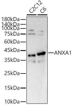 | Western blot analysis of various lysates, using [KO Validated] ANXA1 Rabbit pAb (A1118) at 1:700 dilution. Secondary antibody: HRP-conjugated Goat anti-Rabbit IgG (H+L) (AS014) at 1:10000 dilution. Lysates/proteins: 25μg per lane. Blocking buffer: 3% nonfat dry milk in TBST. Detection: ECL Basic Kit (RM00020). Exposure time: 90s. |
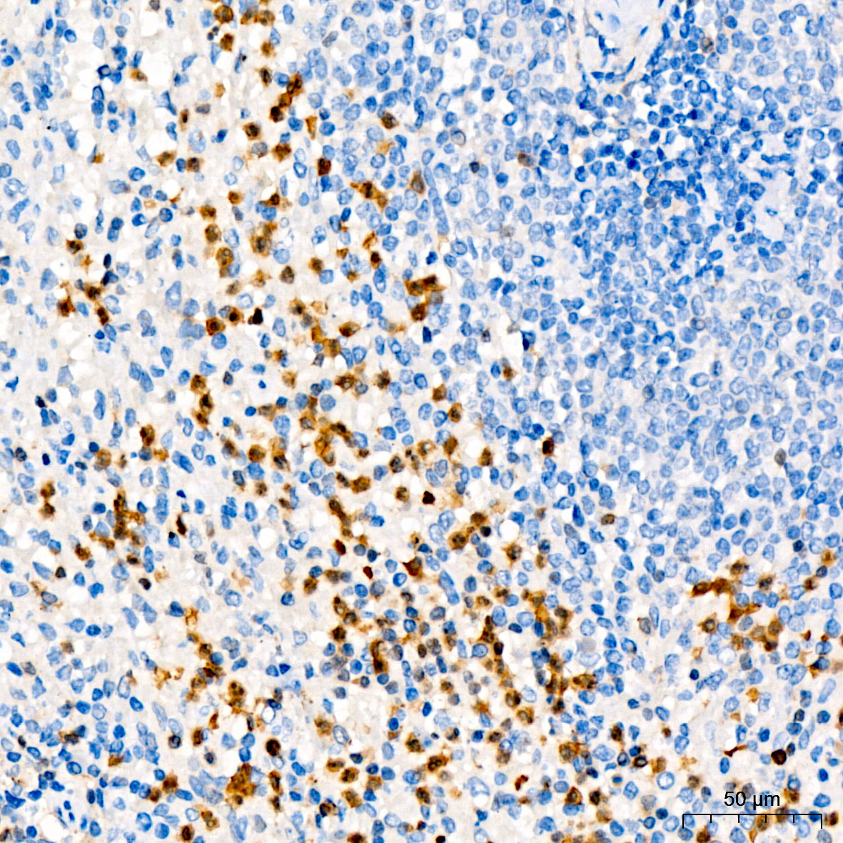 | Immunohistochemistry analysis of paraffin-embedded Human spleen using [KO Validated] ANXA1 Rabbit pAb (A1118) at dilution of 1:50 (40x lens). High pressure antigen retrieval performed with 0.01M Citrate Bufferr (pH 6.0) prior to IHC staining. |
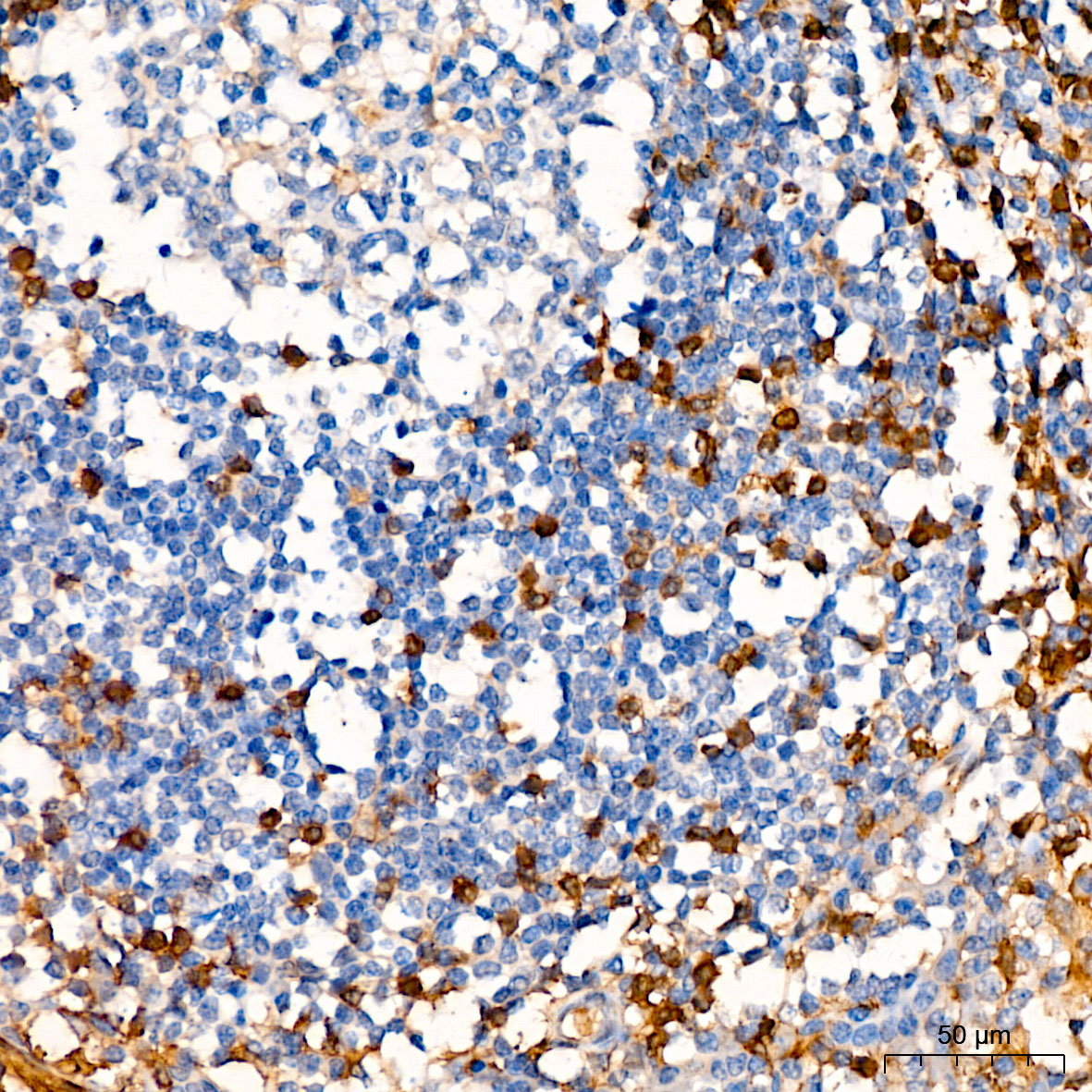 | Immunohistochemistry analysis of paraffin-embedded Human tonsil using [KO Validated] ANXA1 Rabbit pAb (A1118) at dilution of 1:50 (40x lens). High pressure antigen retrieval performed with 0.01M Citrate Bufferr (pH 6.0) prior to IHC staining. |
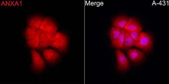 | Immunofluorescence analysis of A-431 cells using [KO Validated] ANXA1 Rabbit pAb (A1118) at dilution of 1:100 (40x lens). Secondary antibody: Cy3-conjugated Goat anti-Rabbit IgG (H+L) (AS007) at 1:500 dilution. Blue: DAPI for nuclear staining. |
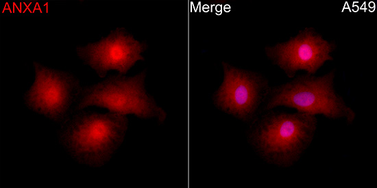 | Immunofluorescence analysis of A-549 cells using [KO Validated] ANXA1 Rabbit pAb (A1118) at dilution of 1:100 (40x lens). Secondary antibody: Cy3-conjugated Goat anti-Rabbit IgG (H+L) (AS007) at 1:500 dilution. Blue: DAPI for nuclear staining. |
 | Immunofluorescence analysis of HeLa cells using [KO Validated] ANXA1 Rabbit pAb (A1118) at dilution of 1:100 (40x lens). Secondary antibody: Cy3-conjugated Goat anti-Rabbit IgG (H+L) (AS007) at 1:500 dilution. Blue: DAPI for nuclear staining. |
You may also be interested in:

