Your shopping cart is empty!
| Reactivity: | Human |
| Applications: | WB, IF/IC, IP, ELISA |
| Host Species: | Rabbit |
| Isotype: | IgG |
| Clonality: | Polyclonal antibody |
| Gene Name: | FUS RNA binding protein |
| Gene Symbol: | FUS |
| Synonyms: | TLS; ALS6; ETM4; FUS1; POMP75; altFUS; HNRNPP2; US |
| Gene ID: | 2521 |
| UniProt ID: | P35637 |
| Immunogen: | Recombinant fusion protein containing a sequence corresponding to amino acids 297-526 of human FUS (NP_004951.1). |
| Dilution: | WB 1:500-1:2000; IF/IC 1:50-1:100 |
| Purification Method: | Affinity purification |
| Concentration: | 0.82 mg/ml |
| Buffer: | PBS with 0.02% sodium azide, 50% glycerol, pH7.3. |
| Storage: | Store at -20°C. Avoid freeze / thaw cycles. |
| Documents: | Manual-FUS polyclonal antibody |
Background
This gene encodes a multifunctional protein component of the heterogeneous nuclear ribonucleoprotein (hnRNP) complex. The hnRNP complex is involved in pre-mRNA splicing and the export of fully processed mRNA to the cytoplasm. This protein belongs to the FET family of RNA-binding proteins which have been implicated in cellular processes that include regulation of gene expression, maintenance of genomic integrity and mRNA/microRNA processing. Alternative splicing results in multiple transcript variants. Defects in this gene result in amyotrophic lateral sclerosis type 6.
Images
 | Western blot analysis of lysates from wild type (WT) and FUS knockout (KO) 293T cells, using [KO Validated] FUS Rabbit pAb (A5921) at 1:3000 dilution. Secondary antibody: HRP-conjugated Goat anti-Rabbit IgG (H+L) (AS014) at 1:10000 dilution. Lysates/proteins: 25μg per lane. Blocking buffer: 3% nonfat dry milk in TBST. Detection: ECL Basic Kit (RM00020). Exposure time: 1s. |
 | Immunofluorescence analysis of C6 cells using [KO Validated] FUS Rabbit pAb (A5921) at dilution of 1:100. Secondary antibody: Cy3-conjugated Goat anti-Rabbit IgG (H+L) (AS007) at 1:500 dilution. Blue: DAPI for nuclear staining. |
 | Immunofluorescence analysis of NIH/3T3 cells using [KO Validated] FUS Rabbit pAb (A5921) at dilution of 1:100. Secondary antibody: Cy3-conjugated Goat anti-Rabbit IgG (H+L) (AS007) at 1:500 dilution. Blue: DAPI for nuclear staining. |
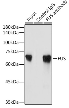 | Immunoprecipitation analysis of 300 μg extracts of Jurkat cells using 3 μg FUS antibody (A5921). Western blot was performed from the immunoprecipitate using FUS antibody (A5921) at a dilution of 1:2000. |
You may also be interested in:

