Your shopping cart is empty!
| Reactivity: | Human |
| Applications: | WB, IF/ICC, ELISA |
| Host Species: | Rabbit |
| Isotype: | IgG |
| Clonality: | Polyclonal antibody |
| Gene Name: | GDP dissociation inhibitor 2 |
| Gene Symbol: | GDI2 |
| Synonyms: | RABGDIB; HEL-S-46e; I2 |
| Gene ID: | 2665 |
| UniProt ID: | P50395 |
| Immunogen: | Recombinant fusion protein containing a sequence corresponding to amino acids 296-445 of human GDI2 (NP_001485.2). |
| Dilution: | WB 1:200-1:3000; IF/IC 1:50-1:100 |
| Purification Method: | Affinity purification |
| Concentration: | 1.26 mg/ml |
| Buffer: | PBS with 0.02% sodium azide, 50% glycerol ,pH7.3. |
| Storage: | Store at -20°C. Avoid freeze / thaw cycles. |
| Documents: | Manual-GDI2 polyclonal antibody |
Background
GDP dissociation inhibitors are proteins that regulate the GDP-GTP exchange reaction of members of the rab family, small GTP-binding proteins of the ras superfamily, that are involved in vesicular trafficking of molecules between cellular organelles. GDIs slow the rate of dissociation of GDP from rab proteins and release GDP from membrane-bound rabs. GDI2 is ubiquitously expressed. The GDI2 gene contains many repetitive elements indicating that it may be prone to inversion/deletion rearrangements. Alternative splicing results in multiple transcript variants encoding distinct isoforms.
Images
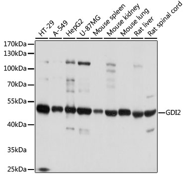 | Western blot analysis of various lysates using [KO Validated] GDI2 Rabbit pAb (A8615) at 1:1000 dilution. Secondary antibody: HRP-conjugated Goat anti-Rabbit IgG (H+L) (AS014) at 1:10000 dilution. Lysates/proteins: 25μg per lane. Blocking buffer: 3% nonfat dry milk in TBST. Detection: ECL Basic Kit (RM00020). Exposure time: 1s. |
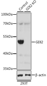 | Western blot analysis of lysates from wild type (WT) and GDI2 knockout (KO) 293T cells, using [KO Validated] GDI2 Rabbit pAb (A8615) at 1:2000 dilution. Secondary antibody: HRP-conjugated Goat anti-Rabbit IgG (H+L) (AS014) at 1:10000 dilution. Lysates/proteins: 25μg per lane. Blocking buffer: 3% nonfat dry milk in TBST. Detection: ECL Basic Kit (RM00020). Exposure time: 1s. |
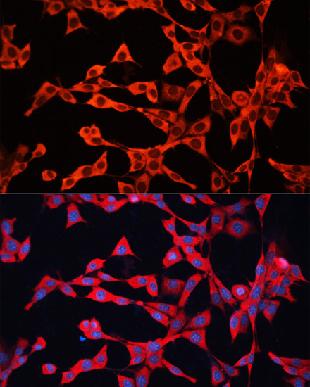 | Immunofluorescence analysis of NIH/3T3 cells using [KO Validated] GDI2 Rabbit pAb (A8615) at dilution of 1:100. Secondary antibody: Cy3-conjugated Goat anti-Rabbit IgG (H+L) (AS007) at 1:500 dilution. Blue: DAPI for nuclear staining. |
You may also be interested in:

