Your shopping cart is empty!
| Reactivity: | Human |
| Applications: | WB, IF/ICC, IP, ELISA |
| Host Species: | Rabbit |
| Isotype: | IgG |
| Clonality: | Polyclonal antibody |
| Gene Name: | G protein subunit alpha i3 |
| Gene Symbol: | GNAI3 |
| Synonyms: | 87U6; HG1A; ARCND1; ARCODS; I3 |
| Gene ID: | 2773 |
| UniProt ID: | P08754 |
| Immunogen: | Recombinant fusion protein containing a sequence corresponding to amino acids 1-354 of human GNAI3 (NP_006487.1). |
| Dilution: | WB 1:500-1:2000; IF/IC 1:50-1:200 |
| Purification Method: | Affinity purification |
| Concentration: | 0.41 mg/ml |
| Buffer: | PBS with 0.02% sodium azide, 50% glycerol ,pH7.3. |
| Storage: | Store at -20°C. Avoid freeze / thaw cycles. |
| Documents: | Manual-GNAI3 polyclonal antibody |
Background
Guanine nucleotide-binding proteins (G proteins) are involved as modulators or transducers in various transmembrane signaling pathways. G proteins are composed of 3 units: alpha, beta and gamma. The gene GNAI3 encodes an alpha subunit and belongs to the G-alpha family. Mutation in this gene, resulting in a gly40-to-arg substitution, is associated with auriculocondylar syndrome, and shown to affect downstream targets in the G protein-coupled endothelin receptor pathway.
Images
 | Western blot analysis of various lysates using [KO Validated] GNAI3 Rabbit pAb (A13307) at 1:1000 dilution. Secondary antibody: HRP-conjugated Goat anti-Rabbit IgG (H+L) (AS014) at 1:10000 dilution. Lysates/proteins: 25μg per lane. Blocking buffer: 3% nonfat dry milk in TBST. Detection: ECL Basic Kit (RM00020). Exposure time: 1s. |
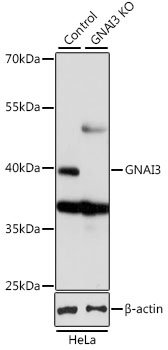 | Western blot analysis of lysates from wild type (WT) and GNAI3 knockout (KO) HeLa cells, using [KO Validated] GNAI3 Rabbit pAb (A13307) at 1:1000 dilution. Secondary antibody: HRP-conjugated Goat anti-Rabbit IgG (H+L) (AS014) at 1:10000 dilution. Lysates/proteins: 25μg per lane. Blocking buffer: 3% nonfat dry milk in TBST. Detection: ECL Basic Kit (RM00020). Exposure time: 1s. |
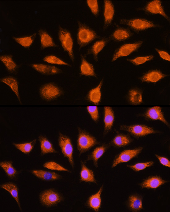 | Immunofluorescence analysis of L929 cells using [KO Validated] GNAI3 Rabbit pAb (A13307) at dilution of 1:100 (40x lens). Secondary antibody: Cy3-conjugated Goat anti-Rabbit IgG (H+L) (AS007) at 1:500 dilution. Blue: DAPI for nuclear staining. |
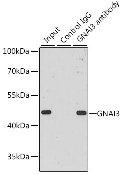 | Immunoprecipitation analysis of 200 μg extracts of MCF-7 cells, using 3 μg GNAI3 antibody (A13307). Western blot was performed from the immunoprecipitate using GNAI3 antibody (A13307) at a dilution of 1:1000. |
You may also be interested in:

