Your shopping cart is empty!
")
KO-Validated LAMP2 Rabbit PolymAb® (20 μl)
| Reactivity: | Human, Mouse, Rat |
| Applications: | WB, IHC, IF/IC, ELISA |
| Host Species: | Rabbit |
| Isotype: | IgG |
| Clonality: | Monoclonal antibody |
| Gene Name: | Lysosomal associated membrane protein 2 |
| Gene Symbol: | LAMP2 |
| Synonyms: | DND; LAMPB; CD107b; LAMP-2; LGP-96; LGP110 |
| Gene ID: | 3920 |
| UniProt ID: | P13473 |
| Immunogen: | Recombinant fusion protein containing a sequence corresponding to amino acids 29-375 of human LAMP2 (NP_002285.1). |
| Dilution: | WB 1:1000-1:6000; IHC 1:500-1:2000; IF/IC 1:200-1:800 |
| Purification Method: | Affinity purification |
| Concentration: | 0.95mg/ml |
| Buffer: | PBS with 0.09% Sodium azide, 0.05% BSA, 50% glycerol, pH7.3. |
| Storage: | Store at -20°C. Avoid freeze / thaw cycles. |
| Documents: | Manual-LAMP2 monoclonal antibody |
Background
The protein encoded by this gene is a member of a family of membrane glycoproteins. This glycoprotein provides selectins with carbohydrate ligands. It may play a role in tumor cell metastasis. It may also function in the protection, maintenance, and adhesion of the lysosome. Alternative splicing of this gene results in multiple transcript variants encoding distinct proteins.
Images
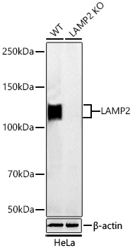 | Western blot analysis of lysates from wild type (WT) and LAMP2 knockout (KO) HeLa cells using [KO Validated] LAMP2 Rabbit PolymAb® (A26781PM) at 1:1000 dilution incubated overnight at 4℃. Secondary antibody: HRP-conjugated Goat anti-Rabbit IgG (H+L) (AS014) at 1:10000 dilution. Lysates/proteins: 25 μg per lane. Blocking buffer: 3% nonfat dry milk in TBST. Detection: ECL Basic Kit (RM00020). Exposure time: 1s. |
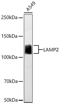 | Western blot analysis of lysates from A-549 cells using [KO Validated] LAMP2 Rabbit PolymAb® (A26781PM) at 1:1000 dilution incubated overnight at 4℃. Secondary antibody: HRP-conjugated Goat anti-Rabbit IgG (H+L) (AS014) at 1:10000 dilution. Lysates/proteins: 25 μg per lane. Blocking buffer: 3% nonfat dry milk in TBST. Detection: ECL Basic Kit (RM00020). Exposure time: 1s. |
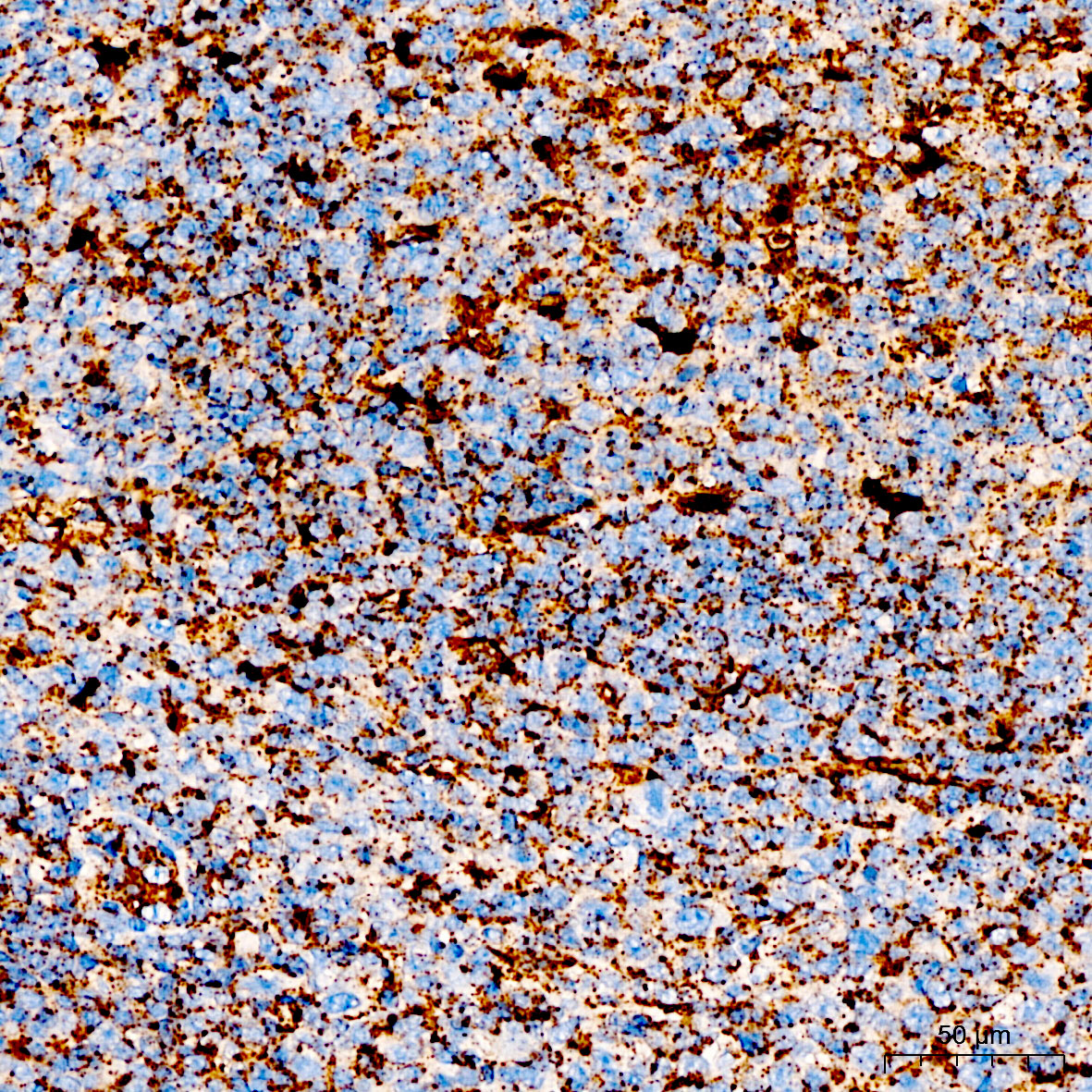 | Immunohistochemistry analysis of paraffin-embedded Human tonsil tissue using [KO Validated] LAMP2 Rabbit PolymAb® (A26781PM) at a dilution of 1:1000 (40x lens). High pressure antigen retrieval performed with 0.01M Citrate Buffer (pH 6.0) prior to IHC staining. |
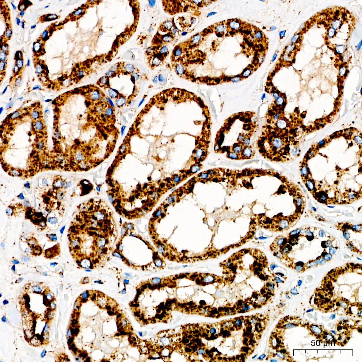 | Immunohistochemistry analysis of paraffin-embedded Human kidney tissue using [KO Validated] LAMP2 Rabbit PolymAb® (A26781PM) at a dilution of 1:1000 (40x lens). High pressure antigen retrieval performed with 0.01M Citrate Buffer (pH 6.0) prior to IHC staining. |
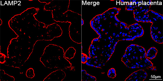 | Confocal imaging of paraffin-embedded Human placenta tissue using [KO Validated] LAMP2 Rabbit PolymAb® (A26781PM, dilution 1:200) followed by a further incubation with Cy3 Goat Anti-Rabbit IgG (H+L) (AS007, dilution 1:500) (Red). DAPI was used for nuclear staining (Blue). High pressure antigen retrieval performed with 0.01M Citrate Buffer (pH 6.0) prior to IF staining. Objective: 40x. |
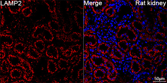 | Confocal imaging of paraffin-embedded Rat kidney tissue using [KO Validated] LAMP2 Rabbit PolymAb® (A26781PM, dilution 1:200) followed by a further incubation with Cy3 Goat Anti-Rabbit IgG (H+L) (AS007, dilution 1:500) (Red). DAPI was used for nuclear staining (Blue). High pressure antigen retrieval performed with 0.01M Citrate Buffer (pH 6.0) prior to IF staining. Objective: 40x. |
You may also be interested in:

