Your shopping cart is empty!
")
KO-Validated Ki67 Rabbit PolymAb® (20 μl)
| Reactivity: | Human, Mouse, Rat |
| Applications: | WB, IHC, IF/IC, ELISA |
| Host Species: | Rabbit |
| Isotype: | IgG |
| Clonality: | Monoclonal antibody |
| Gene Name: | marker of proliferation Ki-67 |
| Gene Symbol: | MKI67 |
| Synonyms: | KIA; MIB-; MIB-1; PPP1R105 |
| Gene ID: | 4288/17345 |
| UniProt ID: | P46013/E9PVX6 |
| Immunogen: | A synthetic peptide corresponding to a sequence within amino acids 2271-2370 of human Ki67 (NP_002408.3). |
| Purification Method: | Affinity purification |
| Concentration: | 0.72mg/ml |
| Buffer: | PBS with 0.09% Sodium azide, 0.05% BSA, 50% glycerol, pH7.3. |
| Storage: | Store at -20°C. Avoid freeze / thaw cycles. |
| Documents: | Manual-MKI67 monoclonal antibody |
Background
Enables protein C-terminus binding activity. Involved in regulation of chromosome segregation and regulation of mitotic nuclear division. Located in chromosome; nuclear body; and nucleolus. Colocalizes with condensed chromosome. Implicated in Crohn's disease; breast cancer; human immunodeficiency virus infectious disease; and pancreatic cancer. Biomarker of several diseases, including Barrett's esophagus; autoimmune disease of musculoskeletal system (multiple); endocrine gland cancer (multiple); gastrointestinal system cancer (multiple); and interstitial cystitis.
Images
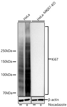 | Western blot analysis of lysates from wild type (WT) and Ki67 knockout (KO) HeLa cells using [KO Validated] Ki67 Rabbit PolymAb® (A26755PM) at 1:1000 dilution incubated overnight at 4℃. HeLa cells and Ki67 knockout (KO) HeLa cells were treated by Nocodazole (1 μg/mL) at 37℃ for 24 hours. Secondary antibody: HRP-conjugated Goat anti-Rabbit IgG (H+L) (AS014) at 1:10000 dilution. Lysates/proteins: 30 μg per lane. Blocking buffer: 3% nonfat dry milk in TBST. Detection: ECL Basic Kit (RM00020). Exposure time: 10s. |
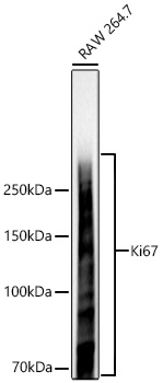 | Western blot analysis of lysates from RAW264.7 cells using [KO Validated] Ki67 Rabbit PolymAb® (A26755PM) at 1:1000 dilution incubated overnight at 4℃. Secondary antibody: HRP-conjugated Goat anti-Rabbit IgG (H+L) (AS014) at 1:10000 dilution. Lysates/proteins: 25 μg per lane. Blocking buffer: 3% nonfat dry milk in TBST. Detection: ECL Basic Kit (RM00020). Exposure time: 5s. |
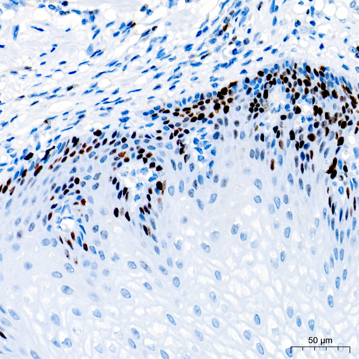 | Immunohistochemistry analysis of paraffin-embedded Human esophagus tissue using [KO Validated] Ki67 Rabbit PolymAb® (A26755PM) at a dilution of 1:1000 (40x lens). High pressure antigen retrieval performed with 0.01M Tris-EDTA Buffer (pH 9.0) prior to IHC staining. |
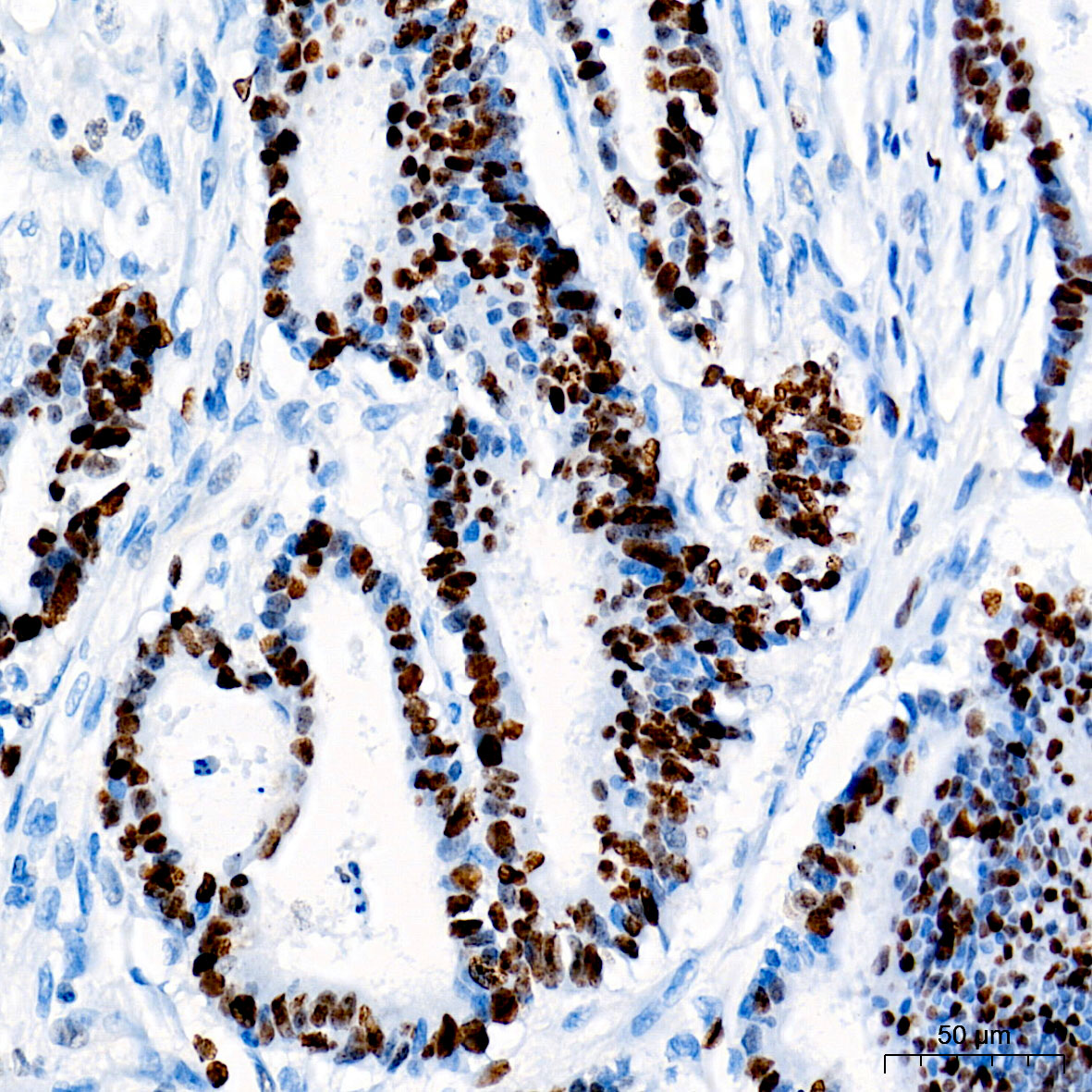 | Immunohistochemistry analysis of paraffin-embedded Human colon carcinoma tissue using [KO Validated] Ki67 Rabbit PolymAb® (A26755PM) at a dilution of 1:1000 (40x lens). High pressure antigen retrieval performed with 0.01M Tris-EDTA Buffer (pH 9.0) prior to IHC staining. |
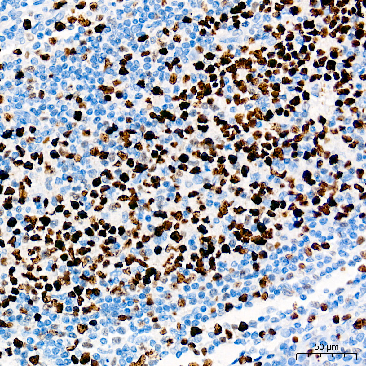 | Immunohistochemistry analysis of paraffin-embedded Human tonsil tissue using [KO Validated] Ki67 Rabbit PolymAb® (A26755PM) at a dilution of 1:1000 (40x lens). High pressure antigen retrieval performed with 0.01M Tris-EDTA Buffer (pH 9.0) prior to IHC staining. |
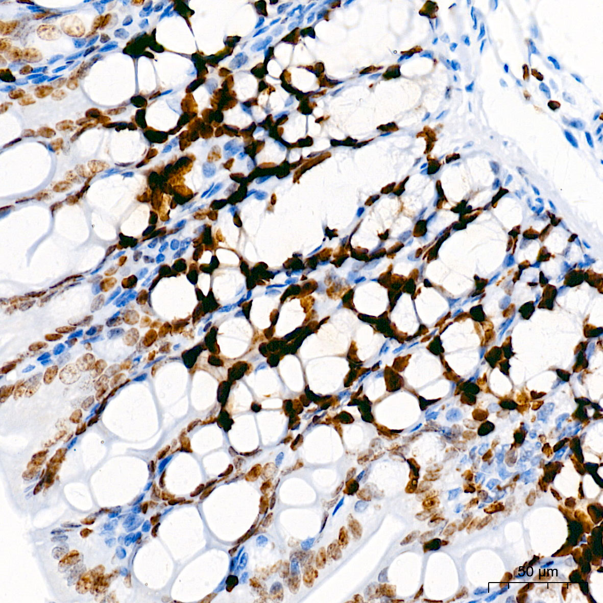 | Immunohistochemistry analysis of paraffin-embedded Mouse colon tissue using [KO Validated] Ki67 Rabbit PolymAb® (A26755PM) at a dilution of 1:1000 (40x lens). High pressure antigen retrieval performed with 0.01M Tris-EDTA Buffer (pH 9.0) prior to IHC staining. |
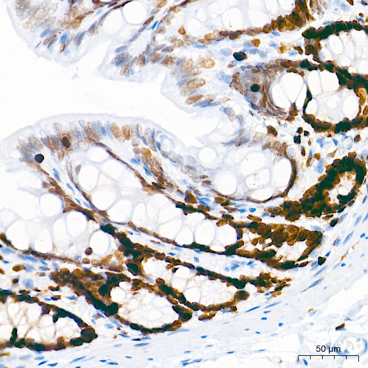 | Immunohistochemistry analysis of paraffin-embedded Rat colon tissue using [KO Validated] Ki67 Rabbit PolymAb® (A26755PM) at a dilution of 1:1000 (40x lens). High pressure antigen retrieval performed with 0.01M Tris-EDTA Buffer (pH 9.0) prior to IHC staining. |
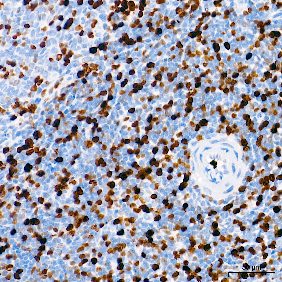 | Immunohistochemistry analysis of paraffin-embedded Rat spleen tissue using [KO Validated] Ki67 Rabbit PolymAb® (A26755PM) at a dilution of 1:1000 (40x lens). High pressure antigen retrieval performed with 0.01M Tris-EDTA Buffer (pH 9.0) prior to IHC staining. |
You may also be interested in:

