Your shopping cart is empty!
")
KO-Validated PEPCK/PCK2 Rabbit mAb (20 μl)
| Reactivity: | Human, Mouse, Rat |
| Applications: | WB, IF/IC, ELISA |
| Host Species: | Rabbit |
| Isotype: | IgG |
| Clonality: | Monoclonal antibody |
| Gene Name: | phosphoenolpyruvate carboxykinase 2, mitochondrial |
| Gene Symbol: | PCK2 |
| Synonyms: | PEPCK; PEPCK2; PEPCK-M; K2 |
| Gene ID: | 5106 |
| UniProt ID: | Q16822 |
| Clone ID: | 10L10J8 |
| Immunogen: | Recombinant fusion protein containing a sequence corresponding to amino acids 550-640 of human PEPCK/PEPCK/PCK2 (Q16822). |
| Dilution: | WB 1:1000-1:4000; IF/IC 1:200-1:800 |
| Purification Method: | Affinity purification |
| Concentration: | 0.5 mg/mL |
| Buffer: | PBS with 0.02% sodium azide, 0.05% BSA, 50% glycerol, pH7.3. |
| Storage: | Store at -20°C. Avoid freeze / thaw cycles. |
| Documents: | Manual-PCK2 monoclonal antibody |
Background
This gene encodes a mitochondrial enzyme that catalyzes the conversion of oxaloacetate to phosphoenolpyruvate in the presence of guanosine triphosphate (GTP). A cytosolic form of this protein is encoded by a different gene and is the key enzyme of gluconeogenesis in the liver. Alternatively spliced transcript variants have been described.
Images
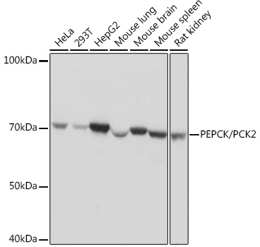 | Western blot analysis of various lysates using PEPCK/PEPCK/PCK2 Rabbit mAb (A4466) at 1:1000 dilution. Secondary antibody: HRP-conjugated Goat anti-Rabbit IgG (H+L) (AS014) at 1:10000 dilution. Lysates/proteins: 25μg per lane. Blocking buffer: 3% nonfat dry milk in TBST. Detection: ECL Basic Kit (RM00020). Exposure time: 1s. |
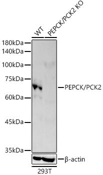 | Western blot analysis of lysates from wild type(WT) and PEPCK/PCK2 knockout (KO) 293T cells, using [KO Validated] PEPCK/PCK2 Rabbit mAb (A4466) at 1:1000 dilution. Secondary antibody: HRP-conjugated Goat anti-Rabbit IgG (H+L) (AS014) at 1:10000 dilution. Lysates/proteins: 25μg per lane. Blocking buffer: 3% nonfat dry milk in TBST. Detection: ECL Basic Kit (RM00020). Exposure time: 30s. |
 | Confocal imaging of HeLa cells using [KO Validated] PEPCK/PCK2 Rabbit mAb (A4466, dilution 1:200) followed by a further incubation with Cy3 Goat Anti-Rabbit IgG (H+L) (AS007, dilution 1:500) (Red). The cells were counterstained with α-Tubulin Mouse mAb (AC012, dilution 1:400) followed by incubation with ABflo® 488-conjugated Goat Anti-Mouse IgG (H+L) Ab (AS076, dilution 1:500) (Green). DAPI was used for nuclear staining (Blue). Objective: 100x. |
 | Confocal imaging of C2C12 cells using [KO Validated] PEPCK/PCK2 Rabbit mAb (A4466, dilution 1:200) followed by a further incubation with Cy3 Goat Anti-Rabbit IgG (H+L) (AS007, dilution 1:500) (Red). The cells were counterstained with α-Tubulin Mouse mAb (AC012, dilution 1:400) followed by incubation with ABflo® 488-conjugated Goat Anti-Mouse IgG (H+L) Ab (AS076, dilution 1:500) (Green). DAPI was used for nuclear staining (Blue). Objective: 100x. |
You may also be interested in:

