Your shopping cart is empty!
")
| Reactivity: | Human, Mouse, Rat |
| Applications: | WB, IF/IC, IP, ELISA |
| Host Species: | Rabbit |
| Isotype: | IgG |
| Clonality: | Monoclonal antibody |
| Gene Name: | Phosphatase and tensin homolog |
| Gene Symbol: | PTEN |
| Synonyms: | BZS; DEC; CWS1; GLM2; MHAM; TEP1; MMAC1; PTEN1; 10q23del; PTENbeta; EN |
| Gene ID: | 5728 |
| UniProt ID: | P60484 |
| Clone ID: | 7O6F4 |
| Immunogen: | Recombinant fusion protein containing a sequence corresponding to amino acids 1-403 of human PTEN (P60484). |
| Dilution: | WB 1:1000-1:2000; IF/IC 1:50-1:200 |
| Purification Method: | Affinity purification |
| Concentration: | 0.6 mg/mL |
| Buffer: | PBS with 0.02% sodium azide, 0.05% BSA, 50% glycerol, pH7.3. |
| Storage: | Store at -20°C. Avoid freeze / thaw cycles. |
| Documents: | Manual-PTEN monoclonal antibody |
Background
This gene was identified as a tumor suppressor that is mutated in a large number of cancers at high frequency. The protein encoded by this gene is a phosphatidylinositol-3,4,5-trisphosphate 3-phosphatase. It contains a tensin like domain as well as a catalytic domain similar to that of the dual specificity protein tyrosine phosphatases. Unlike most of the protein tyrosine phosphatases, this protein preferentially dephosphorylates phosphoinositide substrates. It negatively regulates intracellular levels of phosphatidylinositol-3,4,5-trisphosphate in cells and functions as a tumor suppressor by negatively regulating AKT/PKB signaling pathway. The use of a non-canonical (CUG) upstream initiation site produces a longer isoform that initiates translation with a leucine, and is thought to be preferentially associated with the mitochondrial inner membrane. This longer isoform may help regulate energy metabolism in the mitochondria. A pseudogene of this gene is found on chromosome 9. Alternative splicing and the use of multiple translation start codons results in multiple transcript variants encoding different isoforms.
Images
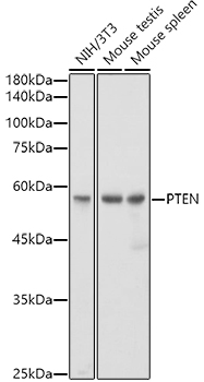 | Western blot analysis of various lysates using [KO Validated] PTEN Rabbit mAb (A19104) at 1:1000 dilution. Secondary antibody: HRP-conjugated Goat anti-Rabbit IgG (H+L) (AS014) at 1:10000 dilution. Lysates/proteins: 25μg per lane. Blocking buffer: 3% nonfat dry milk in TBST. Detection: ECL Basic Kit (RM00020). Exposure time: 1s. |
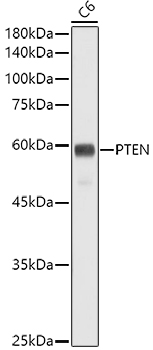 | Western blot analysis of lysates from C6 cells, using [KO Validated] PTEN Rabbit mAb (A19104) at 1:1000 dilution. Secondary antibody: HRP-conjugated Goat anti-Rabbit IgG (H+L) (AS014) at 1:10000 dilution. Lysates/proteins: 25μg per lane. Blocking buffer: 3% nonfat dry milk in TBST. Detection: ECL Basic Kit (RM00020). Exposure time: 60s. |
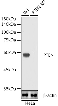 | Western blot analysis of lysates from wild type (WT) and PTEN knockout (KO) HeLa cells, using [KO Validated] PTEN Rabbit mAb (A19104) at 1:1000 dilution. Secondary antibody: HRP-conjugated Goat anti-Rabbit IgG (H+L) (AS014) at 1:10000 dilution. Lysates/proteins: 25μg per lane. Blocking buffer: 3% nonfat dry milk in TBST. Detection: ECL Basic Kit (RM00020). Exposure time: 60s. |
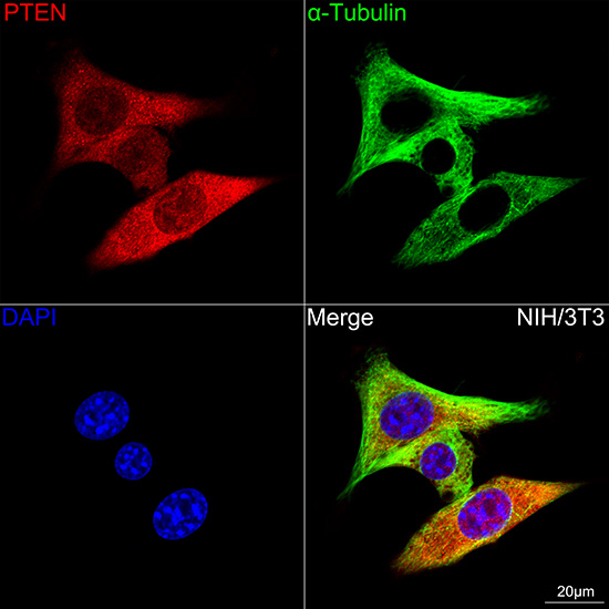 | Confocal imaging of NIH/3T3 cells using [KO Validated] PTEN Rabbit mAb (A19104, dilution 1:100) followed by a further incubation with Cy3 Goat Anti-Rabbit IgG (H+L) (AS007, dilution 1:500) (Red). The cells were counterstained with α-Tubulin Mouse mAb (AC012, dilution 1:400) followed by incubation with ABflo® 488-conjugated Goat Anti-Mouse IgG (H+L) Ab (AS076, dilution 1:500) (Green). DAPI was used for nuclear staining (Blue). Objective: 100x. |
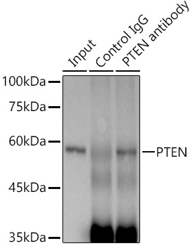 | Immunoprecipitation analysis of 300 μg extracts of NIH/3T3 cells using 3 μg PTEN antibody (A19104). Western blot was performed from the immunoprecipitate using PTEN antibody (A19104) at a dilution of 1:1000. |
You may also be interested in:

