Your shopping cart is empty!
")
KO-Validated N-terminal alpha-synuclein mAb (20 μl)
| Reactivity: | Human, Mouse, Rat |
| Applications: | WB, IF/IC, ELISA |
| Host Species: | Rabbit |
| Isotype: | IgG |
| Clonality: | Monoclonal antibody |
| Gene Name: | Synuclein alpha |
| Gene Symbol: | SNCA |
| Synonyms: | NACP; PARK1; α-Synuclein; SNCA |
| Gene ID: | 6622 |
| UniProt ID: | P37840 |
| Immunogen: | A synthetic peptide corresponding to a sequence corresponding to amino acids 40-50 of human alpha-synuclein(NP_000336.1). |
| Dilution: | WB 1:5000-1:30000; IF/IC 1:200-1:500 |
| Purification Method: | Affinity purification |
| Concentration: | 1.78 mg/ml |
| Buffer: | PBS with 0.09% Sodium azide, 0.05% BSA, 50% glycerol, pH7.3. |
| Storage: | Store at -20°C. Avoid freeze / thaw cycles. |
| Documents: | Manual-SNCA monoclonal antibody |
Background
Alpha-synuclein is a member of the synuclein family, which also includes beta- and gamma-synuclein. Alpha-synuclein is a neuronal protein that plays many roles in synaptic activity such as membrane trafficking of synaptic vesicles and neurotransmitter release. Multiplications in SNCA have been implicated in genetic forms of Parkinson disease and pathological post-translational modifications of alpha-synuclein are a hallmark of Parkinson’s disease. These modifications include phosphorylation, nitration, acetylation, truncation, and aggregation.
Images
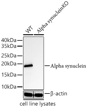 | Western blot analysis of lysates from wild type (WT) and Alpha synuclein knockout (KO) aSyn cells using [KO Validated] N-terminal alpha-synuclein mAb [MJFF-B-3A4] (A27117) at 1:5000 dilution . Secondary antibody: HRP-conjugated Goat anti-Rabbit IgG (H+L) (AS014) at 1:10000 dilution. Lysates/proteins: 25 μg per lane. Blocking buffer: 3% nonfat dry milk in TBST. Detection: ECL Basic Kit (RM00020). Exposure time: 10s. |
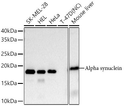 | Western blot analysis of various lysates using [KO Validated] N-terminal alpha-synuclein mAb [MJFF-B-3A4] (A27117) at 1:5000 dilution . Secondary antibody: HRP-conjugated Goat anti-Rabbit IgG (H+L) (AS014) at 1:10000 dilution. Lysates/proteins: 25 μg per lane. Blocking buffer: 3% nonfat dry milk in TBST. Detection: ECL Basic Kit (RM00020). Negative control (NC): T-47D Exposure time: 60s. |
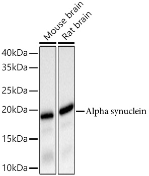 | Western blot analysis of various lysates using [KO Validated] N-terminal alpha-synuclein mAb [MJFF-B-3A4] (A27117) at 1:5000 dilution . Secondary antibody: HRP-conjugated Goat anti-Rabbit IgG (H+L) (AS014) at 1:10000 dilution. Lysates/proteins: 25 μg per lane. Blocking buffer: 3% nonfat dry milk in TBST. Detection: ECL Basic Kit (RM00020). Exposure time: 3s. |
 | Confocal imaging of Mouse primary hippocampal neuron cells(Wild type) and Mouse primary hippocampal neuron cells(SNCA KO) using N-terminal alpha-synuclein mAb [MJFF-B-3A4] (A27117, dilution 1:500) followed by a further incubation with AF488 conjugated donkey Anti-Rabbit (H+L) (A-21206, dilution 1:500) (Green). The cells were counterstained with total aSyn antibody and MAP2 antibody followed by incubation with AF647 conjugated Donkey Anti-mouse IgG (H+L) Ab (A-31571) (Red) and AF568 conjugated Donkey Anti-chicken IgG (H+L) Ab (703-545-155, dilution 1:400) (Grey).DAPI was used for nuclear staining (Blue). Objective: 40x. |
You may also be interested in:

