Your shopping cart is empty!
")
| Reactivity: | Human, Mouse, Rat |
| Applications: | WB, IHC, ELISA |
| Host Species: | Rabbit |
| Isotype: | IgG |
| Clonality: | Monoclonal antibody |
| Gene Name: | signal transducer and activator of transcription 3 |
| Gene Symbol: | STAT3 |
| Synonyms: | APRF; HIES; ADMIO; ADMIO1; T3 |
| Gene ID: | 6774 |
| UniProt ID: | P40763 |
| Immunogen: | A synthetic peptide corresponding to a sequence within amino acids 600-700 of human STAT3 (P40763). |
| Dilution: | WB 1:1000-1:6000; IHC 1:200-1:2000 |
| Purification Method: | Affinity purification |
| Buffer: | PBS with 0.09% Sodium azide, 0.05% BSA, 50% glycerol, pH7.3. |
| Storage: | Store at -20°C. Avoid freeze / thaw cycles. |
| Documents: | Manual-STAT3 monoclonal antibody |
Background
The protein encoded by this gene is a member of the STAT protein family. In response to cytokines and growth factors, STAT family members are phosphorylated by the receptor associated kinases, and then form homo- or heterodimers that translocate to the cell nucleus where they act as transcription activators. This protein is activated through phosphorylation in response to various cytokines and growth factors including IFNs, EGF, IL5, IL6, HGF, LIF and BMP2. This protein mediates the expression of a variety of genes in response to cell stimuli, and thus plays a key role in many cellular processes such as cell growth and apoptosis. The small GTPase Rac1 has been shown to bind and regulate the activity of this protein. PIAS3 protein is a specific inhibitor of this protein. This gene also plays a role in regulating host response to viral and bacterial infections. Mutations in this gene are associated with infantile-onset multisystem autoimmune disease and hyper-immunoglobulin E syndrome.
Images
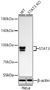 | Western blot analysis of lysates from wild type (WT) and STAT3 knockout (KO) HeLa cells using [KO Validated] STAT3 Rabbit mAb (A19566) at 1:3000 dilution incubated overnight at 4℃. Secondary antibody: HRP-conjugated Goat anti-Rabbit IgG (H+L) (AS014) at 1:10000 dilution. Lysates/proteins: 25 μg per lane. Blocking buffer: 3% nonfat dry milk in TBST. Detection: ECL Basic Kit (RM00020). Exposure time: 30s. |
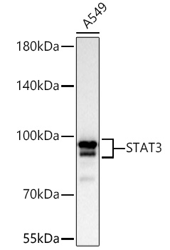 | Western blot analysis of lysates from A549 cells using [KO Validated] STAT3 Rabbit mAb (A19566) at 1:3000 dilution incubated overnight at 4℃. Secondary antibody: HRP-conjugated Goat anti-Rabbit IgG (H+L) (AS014) at 1:10000 dilution. Lysates/proteins: 25 μg per lane. Blocking buffer: 3% nonfat dry milk in TBST. Detection: ECL Basic Kit (RM00020). Exposure time: 30s. |
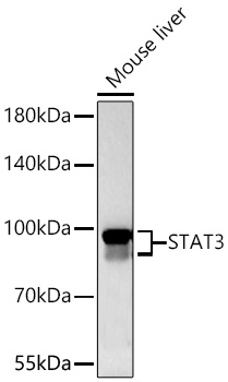 | Western blot analysis of lysates from Mouse liver using [KO Validated] STAT3 Rabbit mAb (A19566) at 1:3000 dilution incubated overnight at 4℃. Secondary antibody: HRP-conjugated Goat anti-Rabbit IgG (H+L) (AS014) at 1:10000 dilution. Lysates/proteins: 25 μg per lane. Blocking buffer: 3% nonfat dry milk in TBST. Detection: ECL Basic Kit (RM00020). Exposure time: 60s. |
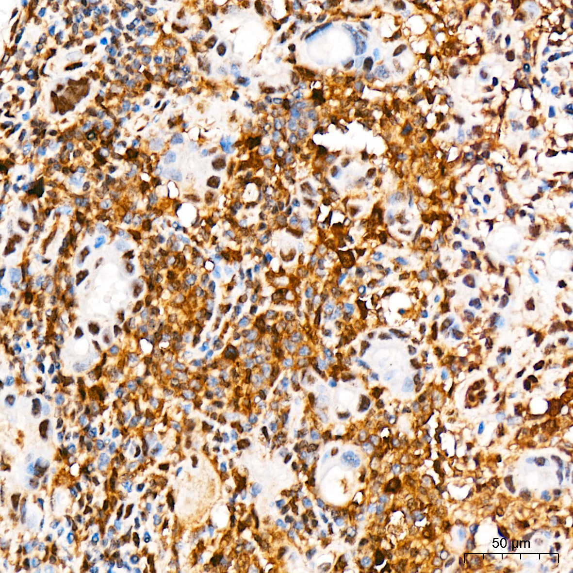 | Immunohistochemistry analysis of paraffin-embedded Human breast tissue using [KO Validated] STAT3 Rabbit mAb (A19566) at a dilution of 1:200 (40x lens). High pressure antigen retrieval was performed with 0.01 M citrate buffer (pH 6.0) prior to IHC staining. |
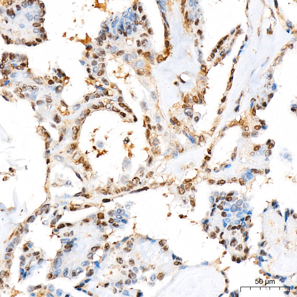 | Immunohistochemistry analysis of paraffin-embedded Human thyroid cancer tissue using [KO Validated] STAT3 Rabbit mAb (A19566) at a dilution of 1:200 (40x lens). High pressure antigen retrieval was performed with 0.01 M citrate buffer (pH 6.0) prior to IHC staining. |
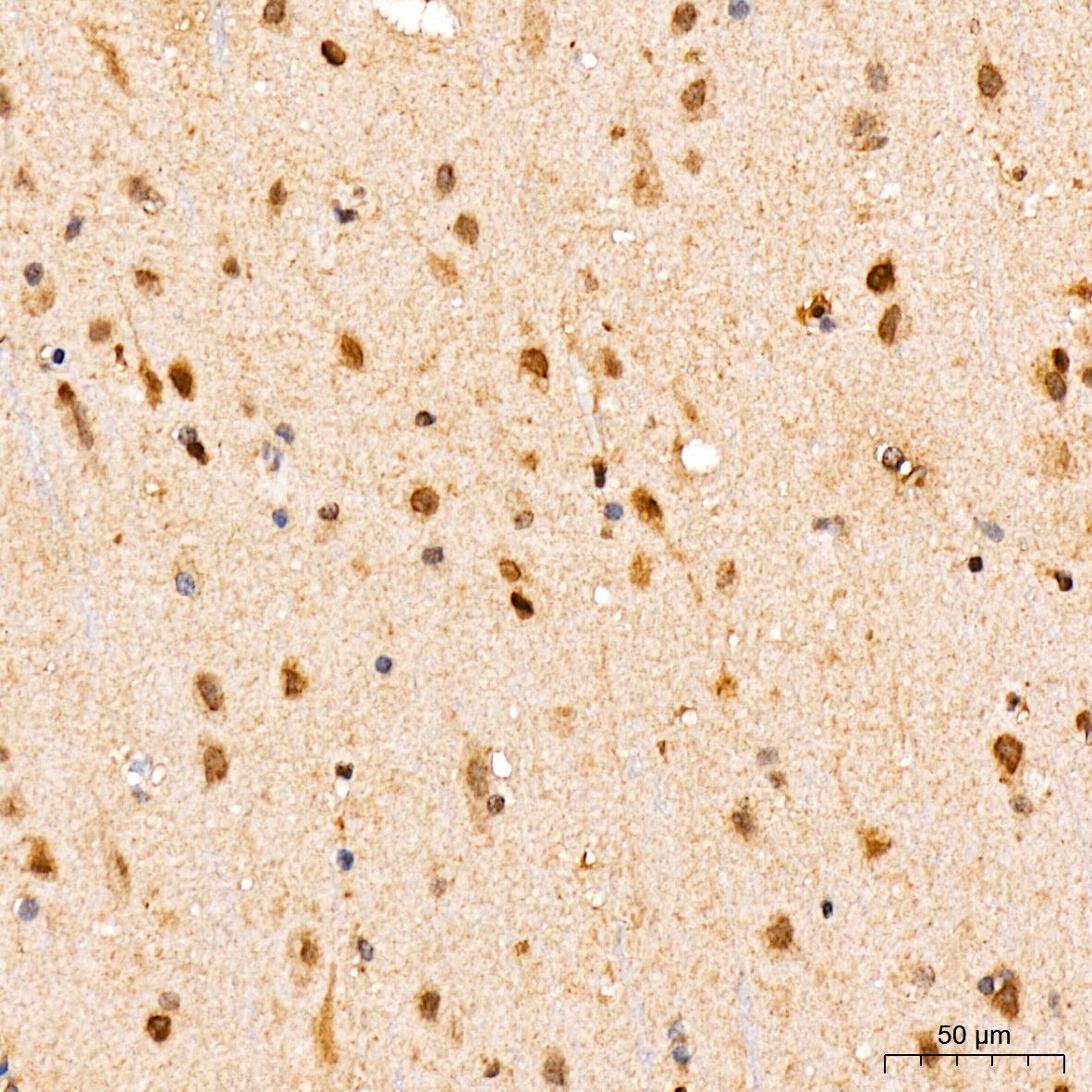 | Immunohistochemistry analysis of paraffin-embedded Human brain tissue using [KO Validated] STAT3 Rabbit mAb (A19566) at a dilution of 1:200 (40x lens). High pressure antigen retrieval was performed with 0.01 M citrate buffer (pH 6.0) prior to IHC staining. |
You may also be interested in:

