eGFP Annexin V and PI Apoptosis Kit is used to detect the externalization of phosphatidylserine in apoptotic cells. This kit provides a sensitive two-color assay that employs eGFP-annexin V and a ready-to-use solution of the red-fluorescent propidium iodide nucleic acid stain. Propidium iodide is impermeant to live cells and apoptotic cells but stains necrotic cells with red fluorescence, binding tightly to the nucleic acids in the cell. After staining a cell population with eGFP-annexin V and propidium iodide in the provided binding buffer, apoptotic cells show green fluorescence, dead cells show red and green fluorescence, and live cells show little or no fluorescence. These populations can easily be distinguished using a flow cytometer with the 488 nm spectral line of an argon-ion laser for excitation.
Specifications
1. Platform: Fluorescence Cytometer
2. Detection Method: Fluorescent
3. Ex/Em: PI: 535/617 nm; eGFP: 475/509 nm
Applications
Cell apoptosis analysis
Components
1. eGFP Annexin V: 500 µl
2. PI: 200 µl
3. 5× Annexin-binding buffer: 50 ml
Storage
Store at 4°C and protect from light.
Case Study
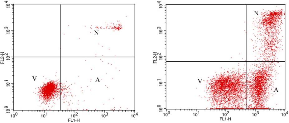
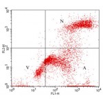
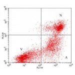
Jurkat cells treated with 10 μM camptothecin for four hours (right panel) or untreated (as control, left panel). Cells were then treated with the reagents in the kit, followed by flow cytometric analysis.
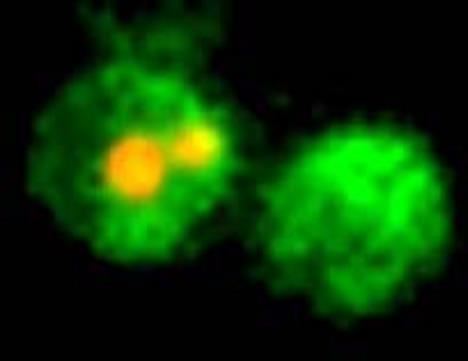
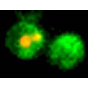
Jurkat cells (T-cell leukemia, human) treated with 10 μM camptothecin for four hours. Cells were then stained with the reagents in the kit. Cells were placed on a glass slide and visualized on a mercury arc lamp microscope. Images were captured on a CCD camera. The right cell is a representative apoptotic cell with only eGFP-Annexin V staining (green plasma membrane), while the left cell is a late stage apoptotic/secondary necrotic cell with both eGFP-Annexin V and PI staining (green membrane with red fragmented nucleus).

