Anti-Myosin light chain 3 Rabbit pAb (100 μl)
| Reactivity: | M,R & M,R & M |
| Applications: | WB & IHC/IF & IEM |
| Host Species: | Rabbit |
| Clonality: | Polyclonal |
| Full Name: | Myosin light chain 3 rabbit polyclonal antibody |
Gene Name: | Myosin light chain 3 |
Synonyms: | Myosin alkali light chain 1, ventricular/slow skeletal muscle isoform, MLC1SB, Myl3, Mlc1v, Mylc, Myosin light chain 3 |
Immunogen: | Recombinant protein corresponding to Mouse Myosin light chain 3 |
Isotype: | IgG |
Purity: | Affinity purification |
Subcellular location: | Cytoplasm |
Predicted MW. | 22 kDa |
Observed MW. | 23 kDa |
Uniprot ID: | P09542, P16409 |
Product Usage Information
Applications | Species | Dilution | Positive tissue |
WB | Mouse, Rat | 1: 500-1: 1000 | heart, muscle |
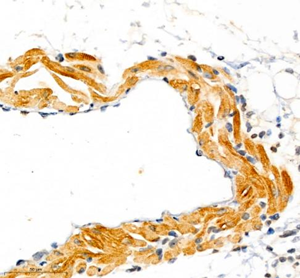
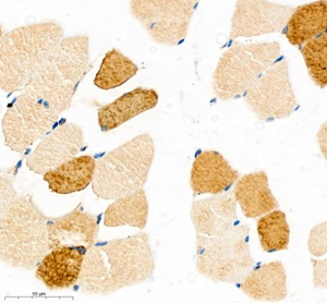
IHC/IF | Mouse, Rat | 1: 1500-1: 3000 | lung, skeletal muscle, heart, esophagus |
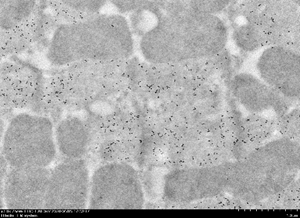
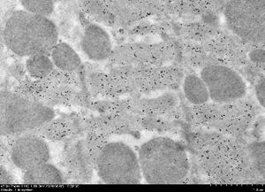
IEM | Mouse | 1: 50 | myocardium |
Background
MYL3, also named as MLC1v, is an essential light chain of myosin that is associated with muscle contraction. It is expressed in ventricular and slow skeletal muscle. MYL3 may serve as a target for caspase-3 in dying cardiomyocytes. Mutations of MYL3 gene cause hypertrophic cardiomyopathy. MYL3 has been identified as potential serum biomarker for drug induced myotoxicity. Great increase in MYL3 serum concentration has been observed in rats with cardiac and skeletal muscle injury.
Images
|
|
Western blot analysis of MYL3 (GB111241) at dilution of 1: 500 |
|
|
Immunohistochemistry of paraffin embedded mouse lung using MYL3 (GB111241) at dilution of 1: 3400 (400x lens) |
|
|
Immunohistochemistry of paraffin embedded mouse skeletal muscle using MYL3 (GB111241) at dilution of 1: 1700 (400x lens) |
|
|
Immunohistochemistry of paraffin embedded rat esophagus using MYL3 (GB111241) at dilution of 1: 3400 (400x lens) |
|
|
Immunohistochemistry of paraffin embedded rat skeletal muscle using MYL3 (GB111241) at dilution of 1: 1700 (400x lens) |
| Immunoelectron microscopy analysis of LR white resin-embedded mouse myocardium using Myl3 (GB111241) at dilution of 1: 50. A goat anti-rabbit antibody preabsorbed with 10nm colloidal gold was used as the secondary antibody, at dilution of 1: 50. |
| Immunoelectron microscopy analysis of LR white resin-embedded mouse myocardium using Myl3 (GB111241) at dilution of 1: 50. A goat anti-rabbit antibody preabsorbed with 10nm colloidal gold was used as the secondary antibody, at dilution of 1: 50. |
| Immunoelectron microscopy analysis of LR white resin-embedded mouse myocardium using Myl3 (GB111241) at dilution of 1: 50. A goat anti-rabbit antibody preabsorbed with 10nm colloidal gold was used as the secondary antibody, at dilution of 1: 50. |
| Immunoelectron microscopy analysis of LR white resin-embedded mouse myocardium using Myl3 (GB111241) at dilution of 1: 50. A goat anti-rabbit antibody preabsorbed with 10nm colloidal gold was used as the secondary antibody, at dilution of 1: 50. |
| Immunoelectron microscopy analysis of LR white resin-embedded mouse myocardium using Myl3 (GB111241) at dilution of 1: 50. A goat anti-rabbit antibody preabsorbed with 10nm colloidal gold was used as the secondary antibody, at dilution of 1: 50. |
| Immunoelectron microscopy analysis of LR white resin-embedded mouse myocardium using Myl3 (GB111241) at dilution of 1: 50. A goat anti-rabbit antibody preabsorbed with 10nm colloidal gold was used as the secondary antibody, at dilution of 1: 50. |
Storage
| Storage | Store at -20°C for one year. Avoid repeated freeze/ thaw cycles. |
| Storage Buffer | PBS with 0.02% sodium azide, 100 μg/ml BSA and 50% glycerol. |


