The LiFluor™ Violet Cell Proliferation Kit offers the convenience of single-use vials for efficient cell labeling. The Violet reagent effortlessly permeates cell membranes and covalently binds to intracellular amines, yielding a stable and enduring fluorescent staining that is compatible with aldehyde-based fixatives. Any surplus unconjugated reagent passively diffuses back into the extracellular medium, where it can be neutralized with complete media and subsequently removed through washing.
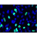
The Violet reagent, post-hydrolysis, exhibits approximate excitation and emission maxima at 405 nm and 450 nm, respectively. Cells marked with the Violet reagent are amenable to visualization under fluorescence microscopy utilizing standard DAPI filter sets or to analysis by flow cytometry on an instrument equipped with a 405 nm excitation source.
Key Features
• Superior performance: Bright, single-peak staining enables visualization of multiple generations.
• Long-term signal stability: Well-retained in cells for several days post stain.
• Easy multiplexing with other fluorophores.
• Simple, robust staining protocol.
Specifications
1. Platform: Flow Cytometer
2. Detection Method: Fluorescent
3. Ex/Em: 405/450 nm
Applications
Cell generation analysis by flow cytometry.
Components
1. Violet: 10 vials
2. Anhydrous DMSO: 500 µl
Storage
Store at -20°C and Protect from light.
Case Study
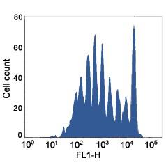
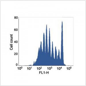
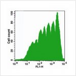
Cell generation analysis with Violet Cell Proliferation Kit. Jurkat cells (~1×10⁶ cells/ml) were stained with Violet dye (1 µM) on Day 0. The cells were cultured for 7 days. Fluorescence intensity was measured with FACS flow cytometer.

