LiFluor™ Cell Proliferation Kits

Tracking Cell Proliferation by Generation after Generation
Our Cell Proliferation Kits can be adapted to multicolor flow cytometry analysis to precisely tracing cell proliferation while performing other cell analysis using different color panel. In addition, the fluorescent labels in our Cell Proliferation Kits can be excited with the most common excitation sources for flow cytometry: the 405, and 488 nm laser, respectively.
| Product Name | Cat. # | Price | View |
| LiFluor™ CFSE Cell Proliferation Kit (1000 rxns) | C0014 | $199 | |
| LiFluor™ Violet Cell Proliferation Kit (200 rxns) | C0015 | $199 |
Tracking New DNA Synthesis
Measuring the synthesis of new DNA is a precise way to assay cell proliferation in individual cells or in cell populations. Our LiFluor™ EdU assays are designed to analyze the rate of new DNA synthesis based on incorporation of the nucleoside analog EdU into DNA. Detection is achieved through a copper-catalyzed “click” reaction that is complete typically within 30 minutes. The LiFluor™ EdU assays have great advantage compared to the traditional BrdU assay. Our LiFluor™ EdU assays use mild reaction conditions, and our bright LiFluor™ 'click' labels offer multicolor choice for multiplexing. The assay kits can be used for cultured cells or tissue sections.
Tracking New RNA Synthesis
Detection of newly synthesized RNA is an important method to measure cell proliferation and toxicological profiling. Our LiFlour™ EU assays utilize an alkyne-modified nucleoside, 5-ethynyl uridine (EU), and powerful click chemistry to detect newly synthesized RNA in a simple, two-step procedure. In step one, the alkyne-containing nucleoside is fed to cells or animals and actively incorporated into nascent RNA. Detection utilizes the “click" reaction between an azide and an alkyne where the modified RNA is detected with a corresponding azide-containing dye.
| Product Name | Cat. # | Price | View |
| LiFluor™ 488 EU Imaging Kit (50 rxns) | C0022 | $349 | |
| LiFluor™ 594 EU Imaging Kit (50 rxns) | C0023 | $349 |
Case Study


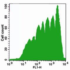
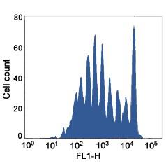
Cell generation analysis with CFSE (Left) and Violet dye (Right). Jurkat cells (~1×10⁶ cells/ml) were stained with CFSE or Violet dye (1 μM) on Day 0. The cells were cultured for 7 days. Fluorescence intensity was measured with FACS flow cytometer.

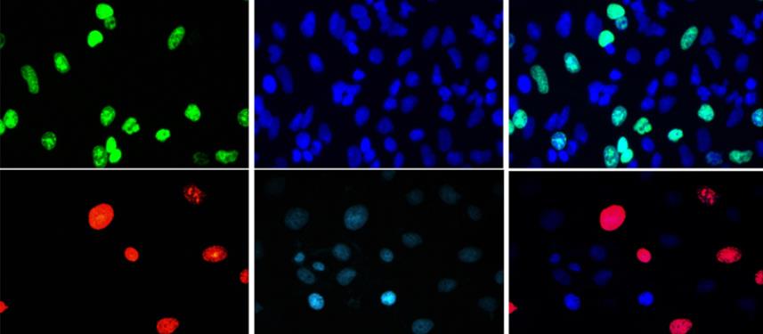
Detection of cell proliferation in cell and mouse tissue. A549 cells were treated with 10 μM EdU for 2 hr, then detected with LiFluor™ 488 azide (green, top), and LiFluor™ 594 azide (red, middle), cells were counterstained with DAPI (blue). A mouse was injected intraperitoneally with 5 mg EdU per kilogram body weight, then sacrificed at 0, 12, and 24 hr.

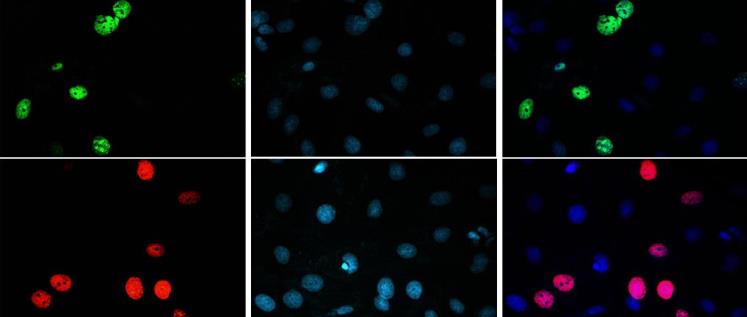
Detection of cell proliferation in cell and mouse tissue. A549 cells were treated with 100 μM EU for 2 hr, then detected with LiFluor™ 488 azide (green, top), and LiFluor™ 594 azide (red, middle), cells were counterstained with DAPI (blue). A mouse was injected intraperitoneally with 50 mg EU per kilogram body weight, then sacrificed at 0, 12, and 24 hr.


